How to recognize complete heart block
- Complete heart block after TAVR, a serious complication, that may first appear as vague symptoms with sudden bradycardia and hypotension.
- Atropine offers limited effectiveness.
- Rapid temporary pacing, close monitoring, and education of patients at high risk for complete heart block can help ensure a good outcome.
History and assessment
Mr. James tells Lizzy, the PACU nurse, that he doesn’t feel well and might pass out. Lizzy knows that conduction disturbances after TAVR can develop hours to days later and has been monitoring cardiac rhythm, HR, and BP. Lizzy notes Mr. James’ slowing heart rate (80 to 40 bpm), and obtains vital signs: BP 82/49 mmHg, RR 22 breaths per minute, and O2 90% on 2 L of O2 by nasal cannula. Mr. James appears pale and diaphoretic. Lizzy recognizes the acute decline as possible heart block. She calls the cardiology NP and obtains a crash cart.
Taking action
The NP orders a 12-lead ECG and asks Lizzy to administer 1 mg of I.V. atropine and prepare for transcutaneous pacing. The ECG reveals complete heart block (CHB) with a ventricular escape rhythm at 38 bpm. As expected in CHB, Mr. James’ heart rate doesn’t increase in response to atropine.
After administering analgesia for comfort, transcutaneous pacing is initiated at 80 bpm. Mr. James’ BP improves to 116/82 mmHg, and he reports feeling better. The interventional cardiologist inserts a transvenous pacemaker, and Mr. James is transferred to the cardiac ICU. Because CHB persists and no other causes are identified, a permanent pacemaker is scheduled for later that day.
Outcome
By postoperative Day 1, Mr. James is ambulating with stable vital signs and no recurrence of dizziness or presyncope. Pacemaker interrogation shows consistent capture with appropriate heart rate response. He’s prepared for discharge home with cardiology follow-up and pacemaker and wound-care education.
Education and follow-up
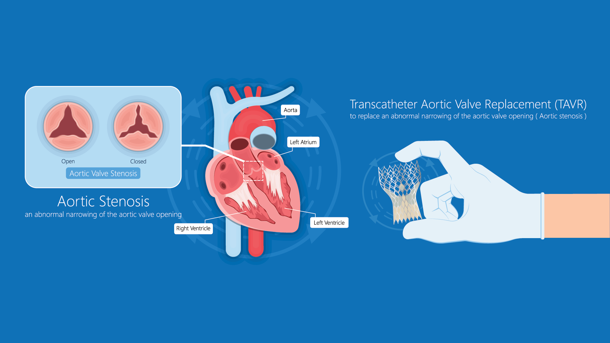
According to Barrett and colleagues, post-TAVR conduction abnormalities result from the proximity of the aortic valve annulus to the heart’s conduction system. During transcatheter valve deployment beneath the AV node and His bundle, mechanical pressure from balloon- or self-expanding prostheses can injure conduction tissue, causing delay or complete heart block. Periprocedural edema and inflammation may lead to delayed conduction disturbances.
According to Barrett and colleagues, CHB incidence after TAVR ranges from 6.5% to 25.9% depending on valve type, implantation depth, and patient-specific risk factors. Rmilah and colleagues note that baseline conduction abnormalities such as right bundle branch block and left anterior fascicular block increase CHB risk. (See Top 5 ECG predictors of complete heart block after TAVR )
Top 5 ECG predictors of complete heart block after TAVR
This table highlights the five highest-yield ECG findings that predict progression to complete heart block (CHB) after trans-
catheter aortic valve replacement (TAVR) and signal the need for closer monitoring and early pacing readiness.
LAFB = left anterior fascicular block, LBBB = left bundle branch block, LPFB = left posterior fascicular block, RBBB = right bundle branch block
Lilly and colleagues recommend up to 14 days of outpatient ambulatory ECG or event monitoring in post-TAVR patients with new or worsening conduction abnormalities if permanent pacing isn’t indicated.
The ACA/AHA/HRS guideline on evaluating and managing bradycardia suggests using atropine for symptomatic bradycardia, although it’s rarely effective because the block often occurs below the atrioventricular node. Anticipate the need for temporary transcutaneous or transvenous pacing. Rapid escalation prevents progression to hemodynamic collapse or cardiac arrest.
The guideline also recommends patient education on recognizing and promptly reporting early symptoms of bradycardia, such as dizziness, syncope, fatigue, and chest discomfort. This case emphasizes the critical role of ongoing nursing assessment to recognize early signs of conduction abnormalities after TAVR.
*Names are fictitious.
Ashley Camp is a cardiology nurse practitioner at Memorial Hermann The Woodlands Medical Center in The Woodlands, Texas.
American Nurse Journal. 2026; 21(8). Doi: 10.51256/ANJ082642
Key words: complete heart block, transcatheter aortic valve replacement, TAVR, bradycardia, transcutaneous pacing