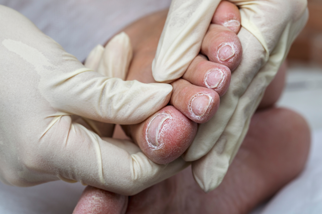
The nurse’s role in assessment, treatment, and education
- People with diabetes face a 10 to 20 times greater risk of amputation because of factors such as peripheral polyneuropathy, poor circulation, and delayed wound healing.
- Diabetic foot ulcers also are associated with significant mortality, with a 5-year mortality rate of 30.5%, which is comparable to the pooled 5-year mortality rate for all cancers.
- Nurses can support better outcomes by educating patients about proper foot care, encouraging self-management, and empowering individuals with diabetes to actively participate in preventing complications and improving quality of life.
Learning Objectives
- Identify key risk factors for diabetic foot complications.
- Understand evidence-based prevention and management strategies.
- Recognize the role of multidisciplinary care in improving outcomes.
Reflective Learning Question 1:
How can recognizing and addressing key risk factors for diabetic foot complications help me intervene earlier and prevent serious outcomes in my patients?
Reflective Learning Question 2:
How does collaborating with a multidisciplinary team enhance patient care and outcomes for individuals at risk of or suffering from diabetic foot complications?
Expiration: 9/1/29
1.5 ANCC contact hours
DIABETIC FOOT COMPLICATIONS represent a significant and growing public health concern worldwide. According to Cho and colleagues and Saeedi and colleagues, diabetes affects 451 million individuals and is expected to increase to nearly 578 million by 2030. Saeedi and colleagues note that over half of people with diabetes are undiagnosed, and 374 million have impaired glucose tolerance. The International Diabetes Federation estimates that 12% of global health expenditure goes toward diabetes, which, according to Cho and colleagues, amounts to about $850 billion. Delayed wound healing, comorbidities, and a proclivity for skin and soft-tissue infections contribute to the significant impact of diabetic foot complications.
These complications arise due to a combination of factors, including peripheral polyneuropathy, poor circulation, and impaired wound healing. In the United States, according to recent epidemiological studies by McDermott and colleagues, diabetic foot ulcers affect an estimated 13% of people with diabetes annually, with 19% to 34% of patients with diabetes developing foot ulcers during their lifetime. Rice and colleagues state that treating a diabetic foot ulcer costs approximately $18,000 per episode, and according to Lipsky and colleagues, diabetic foot ulcers remain the leading cause of nontraumatic lower extremity amputations globally.
Cho and colleagues report that, in addition to the cost and quality-of-life burden, 5 million deaths are attributed to diabetes each year, and the 5-year mortality rate of a diabetic foot ulcer, according to Armstrong and colleagues, is 30.5%, comparable to the 5-year pooled 31% mortality rate of all cancers. It’s not uncommon for diabetic foot ulcers to succumb to the clinical cascade of cellulitis, osteomyelitis, sepsis, amputation, and early mortality. According to studies by Armstrong and colleagues, McDermott and colleagues, among others, individuals with diabetes have a 10 to 20 times higher risk of amputation compared to those without diabetes, with a 20% lifetime incidence of nontraumatic, lower-extremity amputation. Effective management strategies, including regular foot care, early ulcer detection, and multidisciplinary interventions, play a crucial role in reducing the incidence and severity of diabetic foot complications and improving patients’ quality of life.
Glycemic control
Deficient glycemic control is the hallmark of diabetes and the unifying dysfunction causing chronic low-grade inflammation and damage to each body system. Diabetes is associated with mitochondrial dysfunction, which can lead to oxidative stress and the resulting bio-molecular damage that contributes to sterile inflammation (an inflammatory response that occurs without infection, triggered by factors released from damaged or necrotic cells). Hyperglycemia furthers the pro-inflammatory feedback loop, inducing cellular senescence. Hyperglycemia in diabetes and its associated inflammation contribute to systemic damage as a result of impaired cellular metabolism and blood flow.
Immune dysfunction
As described by Appari and colleagues, Barman and Koh, and Berbudi and colleagues, the chronic sterile inflammation that occurs as a result of hyperglycemia is in part due to the immune response, including the pro-inflammatory phenotypical variation of macrophages in fat tissue. Sterile inflammation, according to Berbudi and colleagues, further damages pancreatic beta cells and contributes to insulin deficiencies. Deficient insulin production, with or without insulin resistance, creates a cycle of hyperglycemia and its associated negative effects.
Larsen and colleagues, Pellegrini and colleagues, and Zhang and colleagues note that skin and gut microbiota in adults with diabetes display a distinctive inflammatory profile compared to those without diabetes. Imbalances in the skin and gut microbiome, according to Zhang and colleagues, also contribute to the development and progression of diabetes.
As discussed by Geerlings and Hoepelman and Suaya and colleagues, among others, immune dysfunction in individuals with diabetes can lead to an increased likelihood of developing infection, which may require a lower-limb amputation. Geerlings and Hoepelman note that diabetic cells have shown increased adherence to microorganisms. In a hyperglycemic environment, some microbial species demonstrate increased virulence, making them more likely to cause an infection that becomes severe.
Infected foot ulcer
The photograph shows an infected diabetic foot ulcer to the left heel with necrotic unstable eschar obscuring osteomyelitis. Erythema, edema, and hyperkeratosis are present.
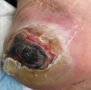
According to Suaya and colleagues, individuals with diabetes who experience hyperglycemia may have deficient pathogen recognition and poor response to infecting microorganisms, leading to increased skin and soft-tissue infections. Nursing interventions that can help protect patients include hand hygiene and education on other infection-prevention techniques, such as wound hygiene. (See Infected foot ulcer.)
Diabetes and wound healing
Multiple pathways (pathophysiological, biochemical, structural, and cellular dysfunction) impair wound healing in patients with diabetes. Hyperglycemia causes increased inflammation in the vasculature, which then triggers cells to become senescent. Senescent cells can’t divide; however, they do secrete more proinflammatory molecules, creating a positive feedback loop in which nonhealing senescent cells create additional senescent cells.
According to Shakeri and colleagues, this process, the senescence-associated secretory phenotype, is more pronounced in diabetes due to hyperglycemia. Wang and colleagues note that the lack of cellular division and the proinflammatory environment may contribute to impaired wound healing as the prevention and removal of senescent cells has been associated with mitigated wound chronicity.
Hyperglycemia impairs the wound-healing process by prolonging inflammation. According to Christman and colleagues, recurring uncontrolled blood glucose levels lead to prolonged time to wound healing. However, Doughty and McNichol and Geerlings and Hoepelman note an association between increased glycemic control and reduced wound complications and improved wound healing.
Elevated hemoglobin A1C levels are associated with delayed wound healing rather than an inability to heal. According to Xiang and colleagues, reasonable blood glucose levels for wound healing are 150 mg/dL to 180 mg/dL or a glycohemoglobin of 7% to 8%.
Vascular disease
Avogaro and colleagues describe diabetic vascular disease as a common comorbidity leading to foot ulceration and amputations. According to Lipsky and colleagues, the disease manifests as peripheral arterial disease (PAD), characterized by narrowed arteries and reduced blood flow to the lower limbs, which impairs wound healing and increases the likelihood of tissue damage. This research indicates that up to 50% of diabetic foot ulcers are associated with PAD, highlighting the critical role of vascular health in diabetic foot care.
Untreated PAD significantly raises the risk of lower-limb amputation. Lipsky and colleagues note that underlying neuropathy and impaired vascular response make patients with diabetes particularly vulnerable to PAD. Early detection of vascular complications via regular screening and prompt intervention, including revascularization procedures when necessary, are essential to improving outcomes and preserving limb function.
Diabetic macrovascular disease affects the large arteries (such as dorsalis pedis and posterior tibial), while microvascular disease affects the small arteries, arterioles, and capillaries. Other vascular problems affected by prolonged and exaggerated hyperglycemia include impaired angiogenesis, red blood cell rigidity, vasoconstriction, hypoperfusion, and contribution to peripheral neuropathy.
Macrovascular impairment largely results from decreasing luminal size. Chronic inflammation and injury from turbid blood flow, leading to plaque formation, causes a reduction in the lumen (the middle of the blood vessel where blood flows). Blood usually flows in a laminar fashion (it stays in the middle of the blood vessel and doesn’t contact the vessel lining). Turbid blood flow is more like a rushing river (it makes contact with the sides of the vessel, causing chronic irritation). Chronic inflammation also leads to microvascular fibrosis (stiffening of the blood vessels), which further limits perfusion.
The final result of altered macro- and microvascular function is decreased skin perfusion. According to Avogaro and colleagues, chronically inflamed endothelium can cause vasoconstriction and further tissue ischemia. Berbudi and colleagues, Feldman and colleagues, and others describe microvascular disease as also strongly associated with peripheral nerve disease, contributing to the development of diabetic neuropathy.
Peripheral neuropathy
Peripheral neuropathy can occur for a variety of reasons in addition to diabetes, including chemotherapy and alcohol use. When it occurs in the context of diabetes it’s called diabetic peripheral neuropathy, which is described as occurring in multiple nerve types and often in multiple body areas. According to Feldman and colleagues, diabetic neuropathy, the most prevalent complication of diabetes, affects more than 50% of patients over the course of the disease. Feldman and colleagues and Martin and colleagues note that glycemic control prevents the onset and progression of diabetic peripheral neuropathy.
In patients with Type 1 diabetes, glycemic control can nearly halt the progression of neuropathy; however, in those with Type 2 diabetes, glycemic control can limit but doesn’t halt its progression. Feldman and colleagues highlight the importance of addressing the additional components of metabolic syndrome in individuals with Type 2 diabetes, including the insulin resistance that can occur in neurons just as it does in fat, muscle, and adipose tissues. Damage to various peripheral nerve types from hyperglycemia causes autonomic, motor, and sensory neuropathies.
Autonomic. As described by Feldman and colleagues, diabetic autonomic neuropathy impairs the sympathetic and parasympathetic nervous system through the involvement of autonomic unmyelinated fibers. According to Sasaki and colleagues, this form of neuropathy also can result in orthostatic hypotension, as well as neurogenic bowel and bladder disorders.
Autonomic nerves usually control sweat and moisture content of the intact skin barrier function; dysfunction can lead to anhidrosis (dry skin). These moisture content alterations compromise the acid mantle of the skin and lead to cracks and fissures, which can serve as portals of entry for pathogens. Berbudi and colleagues note that altered skin barrier function can lead to deficient defense against pathogenic invasion and contribute to increased rates of skin and soft-tissue infection in patients with diabetes.
Sensory. According to Kobayashi and Zochodne, sensory nerves exposed to chronic hyperglycemia undergo atrophy, which in concert with microvascular hypoperfusion results in decreased sensory input and alterations in or loss of sensation. Feldman and colleagues note that sensory neuropathy typically presents in a stocking-and-glove pattern, initially affecting the distal lower extremities where the longest sensory nerve fibers are most susceptible to metabolic and microvascular injury. Symptoms gradually progress proximally and may eventually involve the upper extremities.
Diabetic foot ulcer
The photograph shows a classic presentation of a diabetic foot ulcer beneath the head of the 1st metatarsal. The bony prominence pressure has contributed to a callus and ulcer formation.
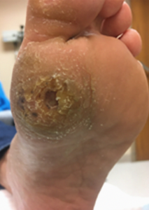
Lack of sensation, particularly in the extremities, can result in ulcer formation and impaired wound healing. Patients with sensory neuropathy don’t receive painful stimuli to their dorsal root ganglia, which would typically trigger avoidance of repetitive trauma to the wound site. Repetitive trauma also can occur in the upper extremities after neuropathy progression. Failure to offload or protect the area appropriately to prevent tissue trauma and associated negative sequelae can lead to ulceration and impaired wound healing. (See Diabetic foot ulcer.)
Motor. The biomolecular neuronal damage related to diabetic neuropathy also targets motor axons, although, as Feldman and colleagues note, to a lesser extent than sensory or autonomic axons. Vink describes how diabetic neuropathy can lead to altered deep tendon reflexes, lower-extremity weakness, and mild muscle wasting.
Motor neuropathy affects the muscles, tendons, and ligaments in the foot. In addition to sensory abnormalities altering gait, motor neuropathy contributes to the formation of foot deformities including hallux valgus, hammertoes, and claw toes. These conditions increase peak pressures over weight-bearing bony prominences, contributing to the formation and progression of diabetic foot ulcers.
Biomechanical complications
Biomechanical diabetic foot issues arise primarily due to peripheral neuropathy and the resulting alterations in foot structure and function. This can result in gait abnormalities, such as increased plantar pressure during walking, which in turn predispose patients to developing diabetic foot ulcers.
Changes in foot biomechanics also contribute to the formation of deformities like Charcot neuroarthropathy (Charcot foot) and toe deformities, further complicating foot function and stability. Effective management of diabetic biomechanical issues involves performing gait assessments, providing orthotic devices to redistribute pressure, and implementing foot care strategies to mitigate the risk of ulceration and structural damage.
Calluses
Diabetic foot calluses, a common manifestation of increased plantar pressure and altered biomechanics in individuals with diabetes, develop due to repetitive friction or pressure, frequently over bony prominences. The calluses are exacerbated by peripheral neuropathy, which reduces pain perception and leads to continued stress on affected areas. If not properly managed, these calluses can progress to diabetic foot ulcers, particularly when accompanied by impaired circulation and compromised wound healing.
Effective management strategies include regular debridement of calluses and appropriate footwear to offload pressure. Patient education regarding foot care practices can help prevent complications.
Foot care practices to prevent and address calluses include managing the skin microclimate with good hygiene followed by appropriate moisturization and footwear. Calluses shouldn’t be managed with pocketknives or rotary tools, which serve as a common source of injury that can lead to diabetic foot infection and ulcer formation. Addressing diabetic foot callus formation early is crucial in preventing more serious sequalae and improving overall foot health. (Learn more about foot assessment and care at myamericannurse.com/foot-assessment-and-care.)
Charcot neuroarthropathy
Charcot neuroarthropathy, a serious limb-threatening and debilitating complication of diabetes, is characterized by progressive joint destruction and deformity in the foot and ankle. It typically occurs in the setting of peripheral neuropathy and frequently presents with swelling, warmth, and redness of the affected limb. Pathophysiology involves neurotraumatic and neurovascular factors that lead to bone resorption and subsequent fractures, further exacerbated by weight-bearing activities and repetitive trauma.
Management of Charcot neuroarthropathy includes early diagnosis via clinical examination and imaging studies, followed by offloading with specialized footwear or total contact casting to prevent further joint damage and ulceration. Timely intervention and ongoing monitoring can help preserve foot function and prevent amputation.
Quality of life
Foot ulcers and complications resulting from diabetes profoundly impact patients’ quality of life, contributing to significant physical, emotional, and social burdens. Studies, including those by Jeffcoate and colleagues, highlight that patients with diabetic foot ulcers experience substantial pain, reduced mobility, and limitations in daily activities, which can lead to increased dependency on caregivers and decreased overall well-being. In addition, the chronic nature of diabetic foot ulcers often necessitates prolonged periods of treatment and frequent medical visits, which have the potential to disrupt normal routines.
Van Netten and colleagues note that these challenges can result in anxiety, depression, and a diminished sense of self-esteem among patients. Effective management and prevention strategies, coupled with comprehensive wound care and psychological support, can help mitigate these impacts and improve the overall quality of life for individuals living with diabetic foot ulcers.
Nursing implications
Multidisciplinary management of diabetic foot complications include the nurse’s role in prevention, assessment, treatment, and patient education. Lipsky and colleagues describe nurses as serving at the forefront of diabetic foot care, conducting regular foot assessments to identify early signs of complications, such as neuropathy, PAD, and skin breakdown. In addition, nurses collaborate closely with podiatrists, endocrinologists, and other healthcare professionals to develop comprehensive care plans tailored to individual patient needs, emphasizing preventive measures such as proper foot hygiene, regular inspection, and appropriate footwear.
Nurses’ crucial role in wound care management includes debridement, provision of advanced therapies, dressing optimization, and monitoring healing progress to prevent development of diabetic foot ulcers and reduce the risk of amputation. Davidson notes that nurses are also key to helping patients achieve ideal glycemic control as an underlying prevention of foot problems. (See Nurse-led patient education)
Nurse-led patient education
Patient self-management education aimed at preventing foot complications in those with diabetes includes the following:
- Clean feet every day with mild soap.
- Dry feet completely.
- To maintain the foot’s skin barrier, moisturize dry areas with lotion.
Foot inspection
- Perform daily checks for sores. Ask a friend or family member to help or use a small mirror or cellphone as needed to enhance visualization.
- If an injury or callus is discovered, contact a provider as soon as possible—diabetic foot problems can escalate quickly.
- Remove shoes at every healthcare appointment for diabetes, not just when seeing a podiatrist.
Footwear
- Check the inside of shoes before donning them. Diabetic neuropathy can cause foot numbness, which may prevent you from feeling objects inside the shoe.
- Wear properly fitting footwear that maintains the skin microclimate (not too wet or dry).
- Shoes should be clean and not worn out. When shoes become worn out, they no longer cushion boney prominences or soles of feet effectively.
- Always wear clean, moisture-wicking socks, such as diabetic socks.
Glycemic control
- Stable blood sugar helps limit the onset and degree of neuropathy and peripheral artery disease.
- Hyperglycemia impairs healing and makes microbes more likely to cause infection.
Home recommendations
- Avoid soaking feet. Soaking can potentially break down skin integrity.
- To avoid the risk of unnoticed trauma, don’t walk barefoot, even at home. Instead, wear custom fit diabetic shoes with inserts, if possible.
- Otherwise, choose footwear with a solid sole and wide toe box that protects feet from trauma.
- Thoroughly clean shared grooming and hygiene tools, such as nail clippers.
Nail care
- Keep toenails trimmed straight across.
- Use an emery board to file sharp edges.
- See a podiatric specialist yearly.
Sources: Swoboda and Held, Swoboda and Fasing
Download Printable Nurse-led patient education Infographic
Empowering patients
Foot complications, common in individuals with diabetes, result from hyperglycemia and its associated chronic inflammation, which contributes to dysfunction of the immune, vascular, and nervous systems. This dysfunction can lead to infection, neuropathy, macro- and microvascular disease, among other co-morbidities. Improved glycemic control can prevent further pathology, reduce persistent multisystem dysfunction, and limit the progression of comorbidities.
Through patient education, nurses empower individuals with diabetes to actively participate in their foot care regimen and promote self-management practices that contribute to improved outcomes, enhanced quality of life, fewer complications, and decreased morbidity.
Laura Swoboda, content expert for this NCPD activity, is a consultant for Convatec, Halyard, Hartmann, Solventum, and Disigns. All relevant financial relationships listed for this individual have been mitigated. None of the other planners or peer reviewers for this educational activity have relevant financial relationship(s) to disclose with ineligible companies whose primary business is producing, marketing, selling, re-selling, or distributing healthcare products used by or on patients.
American Nurse Journal. 2026; 21(9). Doi: 10.51256/ANJ092606
References
Appari M, Channon KM, McNeill E. Metabolic regulation of adipose tissue macrophage function in obesity and diabetes. Antioxid Redox Signal. 2018;29(3):297-312. doi:10.1089/ars.2017.7060
Armstrong DG, Boulton AJM, Bus SA. Diabetic foot ulcers and their recurrence. N Engl J Med. 2017;376(24):2367-75. doi:10.1056/NEJMra1615439
Armstrong DG, Swerdlow MA, Armstrong AA, Conte MS, Padula WV, Bus SA. Five year mortality and direct costs of care for people with diabetic foot complications are comparable to cancer. J Foot Ankle Res. 2020;13(1):16. doi:10.1186/s13047-020-00383-2
Avogaro A, Albiero M, Menegazzo L, de Kreutzenberg S, Fadini GP. Endothelial dysfunction in diabetes: The role of reparatory mechanisms. Diabetes Care. 2011;34(Suppl 2):S285-90. doi:10.2337/dc11-s239
Barman PK, Koh TJ. Macrophage dysregulation and impaired skin wound healing in diabetes. Front Cell Dev Biol. 2020;8:528. doi:10.3389/fcell.2020.00528
Berbudi A, Rahmadika N, Tjahjadi AI, Ruslami R. Type 2 diabetes and its impact on the immune system. Curr Diabetes Rev. 2020;16(5):442-9. doi:10.2174/1573399815666191024085838
Cho NH, Shaw JE, Karuranga S, et al. IDF Diabetes Atlas: Global estimates of diabetes prevalence for 2017 and projections for 2045. Diabetes Res Clin Pract. 2018;138:271-81. doi:10.1016/j.diabres.2018.02.023
Christman AL, Selvin E, Margolis DJ, Lazarus GS, Garza LA. Hemoglobin A1c predicts healing rate in diabetic wounds. J Invest Dermatol. 2011;131(10):2121-7. doi:10.1038/jid.2011.176
Davidson MB. Effect of diabetes-trained nurse practitioners on glycemic outcomes: Their suggested use in busy primary care practices. Clin Diabetes. 2021;39(3):293-6. doi:10.2337/cd20-0102
Doughty DB, McNichol L. Wound, Ostomy and Continence Nurses Society® Core Curriculum: Wound Management. Philadelphia, PA: Wolters Kluwer; 2021.
Dryden M, Baguneid M, Eckmann C, et al. Pathophysiology and burden of infection in patients with diabetes mellitus and peripheral vascular disease: Focus on skin and soft-tissue infections. Clin Microbiol Infect. 2015;21(Suppl 2):S27-32. doi:10.1016/j.cmi.2015.03.024
Feldman EL, Callaghan BC, Pop-Busui R, et al. Diabetic neuropathy. Nat Rev Dis Primers. 2019;5(1):41. doi:10.1038/s41572-019-0092-1
Geerlings SE, Hoepelman AI. Immune dysfunction in patients with diabetes mellitus (DM). FEMS Immunol Med Microbiol. 1999;26(3-4):259-65. doi:10.1111/j.1574-695X.1999.tb01397.x
International Diabetes Federation. IDF Diabetes Atlas 8th edition. 2017 fmdiabetes.org/wp-content/uploads/2018/03/IDF-2017.pdf
Jeffcoate WJ, Vileikyte L, Boyko EJ, Armstrong DG, Boulton AJM. Current challenges and opportunities in the prevention and management of diabetic foot ulcers. Diabetes Care. 2018;41(4):645-52. doi:10.2337/dc17-1836
Jenkins TC, Knepper BC, Jason Moore S, et al. Comparison of the microbiology and antibiotic treatment among diabetic and nondiabetic patients hospitalized for cellulitis or cutaneous abscess. J Hosp Med. 2014;9(12):788-94. doi:10.1002/jhm.2267
Kobayashi M, Zochodne DW. Diabetic neuropathy and the sensory neuron: New aspects of pathogenesis and their treatment implications. J Diabetes Investig. 2018;9(6):1239-54. doi:10.1111/jdi.12833
Larsen N, Vogensen FK, van den Berg FWJ, et al. Gut microbiota in human adults with type 2 diabetes differs from non-diabetic adults. PLoS One. 2010;5(2):e9085. doi:10.1371/journal.pone.0009085
Lipsky BA, Senneville É, Abbas ZG, et al. Guidelines on the diagnosis and treatment of foot infection in persons with diabetes (IWGDF 2019 update). Diabetes Metab Res Rev. 2020;36(Suppl 1):e3280. doi:10.1002/dmrr.3280
Martin CL, Albers JW, Pop-Busui R. Neuropathy and related findings in the diabetes control and complications trial/epidemiology of diabetes interventions and complications study. Diabetes Care. 2014;37(1):31-8. doi: 10.2337/dc13-2114
McDermott K, Fang M, Boulton AJM, Selvin E, Hicks CW. Etiology, epidemiology, and disparities in the burden of diabetic foot ulcers. Diabetes Care. 2023;46 (1): 209-21. doi:10.2337/dci22-0043
Pellegrini S, Sordi V, Bolla AM, et al. Duodenal mucosa of patients with type 1 diabetes shows distinctive inflammatory profile and microbiota. J Clin Endocrinol Metab. 2017;102(5):1468-77. doi:10.1210/jc.2016-3222
Regulski M. Understanding diabetic induction of cellular senescence: A concise review. Wounds. 2018;30(4):96-101.
Rice JB, Desai U, Ristovska L, et al. Economic outcomes among Medicare patients receiving bioengineered cellular technologies for treatment of diabetic foot ulcers. J Med Econ. 2015;18(8):586-95. doi:10.3111/13696998.2015.1031793
Saeedi P, Petersohn I, Salpea P, et al. Global and regional diabetes prevalence estimates for 2019 and projections for 2030 and 2045: Results from the International Diabetes Federation Diabetes Atlas, 9th edition. Diabetes Res Clin Pract. 2019;157:107843. doi:10.1016/j.diabres.2019.107843
Sasaki H, Kawamura N, Dyck PJ, Dyck PJB, Kihara M, Low PA. Spectrum of diabetic neuropathies. Diabetol Int. 2020;11(2):87-96. doi:10.1007/s13340-019-00424-7
Shakeri H, Lemmens K, Gevaert AB, De Meyer GRY, Segers VFM. Cellular senescence links aging and diabetes in cardiovascular disease. Am J Physiol Heart Circ Physiol. 2018;315(3):H448-62. doi:10.1152/ajpheart.00287.2018
Suaya JA, Eisenberg DF, Fang C, Miller LG. Skin and soft tissue infections and associated complications among commercially insured patients aged 0-64 years with and without diabetes in the U.S. PLoS One. 2013;8(4): e60057. doi:10.1371/journal.pone.0060057
Swoboda L, Bell D, Bauer K, Clayton S. Time out before amputation. Adv Skin Wound Care. 2026;39(5):234-5. doi:10.1097/ASW.0000000000000450
Swoboda L, Fasing S. Getting ready for certification: Nail debridement. J Wound Ostomy Continence Nurs. 2024;51(2):156-8. doi:10.1097/WON.0000000000001068
Swoboda L, Held J. Impaired wound healing in diabetes. J Wound Care. 2022;31(10):882-5. doi:10.12968/jowc.2022.31.10.882
van Netten JJ, Bus SA, Apelqvist J, et al. Definitions and criteria for diabetic foot disease. Diabetes Metab Res Rev. 2020;36(Suppl 1):e3268. doi:10.1002/dmrr.3268
Vinik AI. Diabetic sensory and motor neuropathy [published corrections appear in N Engl J Med. 2016;374(18):1797, and N Engl J Med. 2016;375(14 ):1402]. N Engl J Med. 2016;374(15):1455-64. doi:10.1056/NEJMcp1503948
Wang Z, Chen Z, Jiang Z, et al. Cordycepin prevents radiation ulcer by inhibiting cell senescence via NRF2 and AMPK in rodents. Nat Commun. 2019;10(1):2538. doi:10.1038/s41467-019-10386-8
Wang Z, Shi C. Cellular senescence is a promising target for chronic wounds: A comprehensive review. Burns Trauma. 2020;8:tkaa021. doi:10.1093/burnst/tkaa021
Wang H, Wang Z, Huang Y, et al. Senolytics (DQ) mitigates radiation ulcers by removing senescent cells. Front Oncol. 2020;9:1576. doi:10.3389/fonc.2019.01576
Xiang J, Wang S, He Y, Xu L, Zhang S, Tang Z. Reasonable glycemic control would help wound healing during the treatment of diabetic foot ulcers. Diabetes Ther. 2019;10(1):95-105. doi:10.1007/s13300-018-0536-8
Zhang S, Cai Y, Meng C, et al. The role of the microbiome in diabetes mellitus. Diabetes Res Clin Pract. 2021;172:108645. doi:10.1016/j.diabres.2020.108645
Key words: diabetes, foot care, diabetic foot ulcer, podiatry