Respiratory failure is one of the most common reasons for admission to the intensive care unit (ICU) and a common comorbidity in patients admitted for acute care. What’s more, it’s the leading cause of death from pneumonia and chronic obstructive pulmonary disease (COPD) in the United States. This article briefly reviews the physiologic components of respiration, differentiates the main types of respiratory failure, and discusses medical treatment and nursing care for patients with respiratory failure.
Physiologic components of ventilation and respiration
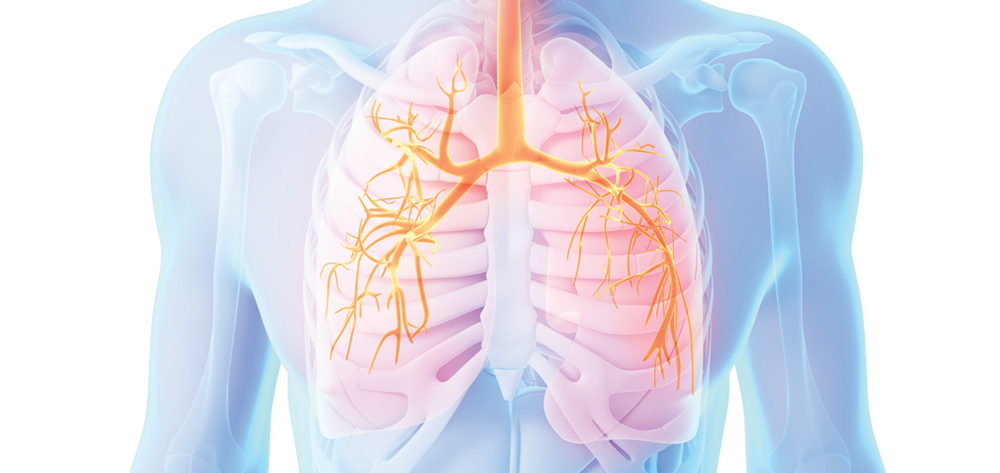
The lung is highly elastic. Lung inflation results from the partial pressure of inhaled gases and the diffusion-pressure gradient of these gases across the alveolar-capillary membrane. The lungs play a passive role in breathing, but ventilation requires muscular effort. When the diaphragm contracts, the thoracic cavity enlarges, causing the lungs to inflate. During forced inspiration when a large volume of air is inspired, external intercostal muscles act as a second set of inspiratory muscles.
Accessory muscles in the neck and chest are the last group of inspiratory muscles, used only for deep and heavy breathing, such as during intense exercise or respiratory failure. During expiration, the diaphragm relaxes, decreasing thoracic cavity size and causing the lungs to deflate. With normal breathing, expiration is purely passive. But with exercise or forced expiration, expiratory muscles (including the abdominal wall and internal intercostal muscles) become active. These important muscles are necessary for coughing.
Respiration—the process of exchanging oxygen (O2) and carbon dioxide (CO2)—involves ventilation, oxygenation, and gas transport; the ventilation/perfusion (V/Q) relationship; and control of breathing. Respiration is regulated by chemical and neural control systems, including the brainstem, peripheral and central chemoreceptors, and mechanoreceptors in skeletal muscle and joints. (See Control of breathing.)
A dynamic process, ventilation is affected by the respiratory rate (RR) and tidal volume—the amount of air inhaled and exhaled with each breath. Pulmonary ventilation refers to the total volume of air inspired or expired per minute.
Not all inspired air participates in gas exchange. Alveolar ventilation—the volume of air entering alveoli taking part in gas exchange—is the most important variable in gas exchange. Air that distributes to the conducting airways is deemed dead space or wasted air because it’s not involved in gas exchange. (See Oxygenation and gas transport.)
Ultimately, effective ventilation is measured by the partial pressure of CO2 in arterial blood (Paco2). All expired CO2 comes from alveolar gas. During normal breathing, the breathing rate or depth adjusts to maintain a steady Paco2 between 35 and 45 mm Hg. Hyperventilation manifests as a low Paco2; hypoventilation, as a high Paco2. During exercise or certain disease states, increasing breathing depth is far more effective than increasing the RR in improving alveolar ventilation.
Lung recoil and compliance
The lungs, airways, and vascular trees are embedded in elastic tissue. To inflate, the lung must stretch to overcome these elastic components. Elastic recoil—the lung’s ability to return to its original shape after stretching from inhalation—relates inversely to compliance. Lung compliance indirectly reflects lung stiffness or resistance to stretch. A stiff lung, as in pulmonary fibrosis, is less compliant than a normal lung.
With reduced compliance, more work is required to produce a normal tidal volume. With extremely high compliance, as in emphysema where there is loss of alveolar and elastic tissue, the lungs inflate extremely easily. Someone with emphysema must expend a lot of effort to get air out of the lungs because they don’t recoil back to their normal position during expiration. In both pulmonary fibrosis and emphysema, inadequate lung ventilation leads to hypercapnic respiratory failure.
Respiratory failure
Respiratory failure occurs when one of the gas-exchange functions—oxygenation or CO2 elimination—fails. A wide range of conditions can lead to acute respiratory failure, including drug overdose, respiratory infection, and exacerbation of chronic respiratory or cardiac disease.
Respiratory failure may be acute or chronic. In acute failure, life-threatening derangements in arterial blood gases (ABGs) and acid-base status occur, and patients may need immediate intubation. Respiratory failure also may be classified as hypoxemic or hypercapnic.
Clinical indicators of acute respiratory failure include:
- partial pressure of arterial oxygen (Pao2) below 60 mm Hg, or arterial oxygen saturation as measured by pulse oximetry (Spo2) below 91% on room air
- Paco2 above 50 mm Hg and pH below 7.35
- Pao2 decrease or Paco2 increase of 10 mm Hg from baseline in patients with chronic lung disease (who tend to have higher Paco2 and lower PaO2 baseline values than other patients).
In contrast, chronic respiratory failure is a long-term condition that develops over time, such as with COPD. Manifestations of chronic respiratory failure are less dramatic and less apparent than those of acute failure.
Three main types of respiratory failure
The most common type of respiratory failure is type 1, or hypoxemic respiratory failure (failure to exchange oxygen), indicated by a Pao2 value below 60 mm Hg with a normal or low Paco2 value. In ICU patients, the most common causes of type 1 respiratory failure are V/Q mismatching and shunts. COPD exacerbation is a classic example of V/Q mismatching. Shunting, which occurs in virtually all acute lung diseases, involves alveolar collapse or fluid-filled alveoli. Examples of type 1 respiratory failure include pulmonary edema (both cardiogenic and noncardiogenic), pneumonia, influenza, and pulmonary hemorrhage. (See Ventilation and perfusion: A critical relationship.)
Type 2, or hypercapnic, respiratory failure, is defined as failure to exchange or remove CO2, indicated by Paco2 above 50 mm Hg. Patients with type 2 respiratory failure who are breathing room air commonly have hypoxemia. Blood pH depends on the bicarbonate level, which is influenced by hypercapnia duration. Any disease that affects alveolar ventilation can result in type 2 respiratory failure. Common causes include severe airway disorders (such as COPD), drug overdose, chest-wall abnormalities, and neuromuscular disease.
Type 3 respiratory failure (also called perioperative respiratory failure) is a subtype of type 1 and results from lung or alveolar atelectasis. General anesthesia can cause collapse of dependent lung alveoli. Patients most at risk for type 3 respiratory failure are those with chronic lung conditions, excessive airway secretions, obesity, immobility, and tobacco use, as well as those who’ve had surgery involving the upper abdomen. Type 3 respiratory failure also may occur in patients experiencing shock, from hypoperfusion of respiratory muscles. Normally, less than 5% of total cardiac output flows to respiratory muscles. But in pulmonary edema, lactic acidosis, and anemia (conditions that commonly arise during shock), up to 40% of cardiac output may flow to the respiratory muscles.
Signs and symptoms of respiratory failure
Patients with impending respiratory failure typically develop shortness of breath and mental-status changes, which may present as anxiety, tachypnea, and decreased Spo2 despite increasing amounts of supplemental oxygen.
Acute respiratory failure may cause tachycardia and tachypnea. Other signs and symptoms include periorbital or circumoral cyanosis, diaphoresis, accessory muscle use, diminished lung sounds, inability to speak in full sentences, an impending sense of doom, and an altered mental status. The patient may assume the tripod position in an attempt to further expand the chest during the inspiratory phase of respiration. In chronic respiratory failure, the only consistent clinical indictor is protracted shortness of breath.
Be aware that pulse oximetry measures the percentage of hemoglobin saturated with oxygen, but it doesn’t give information about oxygen delivery to the tissues or the patient’s ventilatory function. So be sure to consider the patient’s entire clinical presentation. Compared to SpO2, an ABG study provides more accurate information on acid-base balance and blood oxygen saturation. Capnography is another tool used for monitoring patients receiving anesthesia and in critical care units to assess a patient’s respiratory status. It directly monitors inhaled and exhaled concentration of CO2 and indirectly monitors Paco2.
Treatment and management
In acute respiratory failure, the healthcare team treats the underlying cause while supporting the patient’s respiratory status with supplemental oxygen, mechanical ventilation, and oxygen saturation monitoring. Treatment of the underlying cause, such as pneumonia, COPD, or heart failure, may require diligent administration of antibiotics, diuretics, steroids, nebulizer treatments, and supplemental O2 as appropriate.
For chronic respiratory failure, despite the wide range of chronic or end-stage pathology present (such as COPD, heart failure, or systemic lupus erythematosus with lung involvement), the mainstay of treatment is continuous supplemental O2, along with treatment of the underlying cause.
Nursing care
Nursing care can have a tremendous impact in improving efficiency of the patient’s respiration and ventilation and increasing the chance for recovery. To detect changes in respiratory status early, assess the patient’s tissue oxygenation status regularly. Evaluate ABG results and indices of end-organ perfusion. Keep in mind that the brain is extremely sensitive to O2 supply; decreased O2 can lead to an altered mental status. Also, know that angina signals inadequate coronary artery perfusion. In addition, stay alert for conditions that can impair O2 delivery, such as elevated temperature, anemia, impaired cardiac output, acidosis, and sepsis.
As indicated, take steps to improve V/Q matching, which is crucial for improving respiratory efficiency. To enhance V/Q matching, turn the patient on a regular and timely basis to rotate and maximize lung zones. Because blood flow and ventilation are distributed preferentially to dependent lung zones, V/Q is maximized on the side on which the patient is lying.
Regular, effective use of incentive spirometry helps maximize diffusion and alveolar surface area and can help prevent atelectasis. Regular rotation of V/Q lung zones by patient turning and repositioning enhances diffusion by promoting a healthy, well-perfused alveolar surface. These actions, as well as suctioning, help mobilize sputum or secretions.
Nutritional support
Patients in respiratory failure have unique nutritional needs and considerations. Those with acute respiratory failure from primary lung disease may be malnourished initially or may become malnourished from increased metabolic demands or inadequate nutritional intake. Malnutrition can impair the function of respiratory muscles, reduce ventilatory drive, and decrease lung defense mechanisms. Clinicians should consider nutritional support and individualize such support to ensure adequate caloric and protein intake to meet the patient’s respiratory needs.
Patient and family education
Provide appropriate education to the patient and family to promote adherence with treatment and help prevent the need for readmission. Explain the purpose of nursing measures, such as turning and incentive spirometry, as well as medications. At discharge, teach patients about pertinent risk factors for their specific respiratory condition, when to return to the healthcare provider for follow-up care, and home measures they can take to promote and maximize respiratory function.
Selected references
Cooke CR, Erikson SE, Eisner MD, Martin GS. Trends in the incidence of noncardiogenic acute respiratory failure: the role of race. Crit Care Med. 2012;40(5):1532-8.
Gehlbach BK, Hall JB. Respiratory failure and mechanical ventilation. In Porter RS, ed. The Merck Manual. 19th ed. West Point, PA; Merck Sharp & Dohme Corp.; 2011.
Kaynar AM. Respiratory failure treatment and management. Updated August 14, 2014. http://emedicine.medscape.com/article/167981-treatment#aw2aab6b6b2. Accessed August 23, 2014.
Kress JP, Hall JB: Approach to the patient with critical illness. In Longo DL, Fauci AS, Kasper DL, et al., eds. Harrison’s Principles of Internal Medicine. 18th ed. New York, NY: McGraw-Hill Professional; 2012.
Pinson R. Revisiting respiratory failure, part 1. ACP Hosp. 2013;Oct:5-6. www.acphospitalist.org/archives/2013/10/coding.htm. Accessed August 23, 2014.
Pinson R. Revisiting respiratory failure, part 2. ACP Hosp. 2013;Nov:7-8. www.acphospitalist.org/archives/2013/11/coding.htm. Accessed October 3, 2014.
Schraufnagel D. Breathing in America: Diseases, Progress, and Hope. American Thoracic Society; 2010.
Michelle Fournier is director of clinical consulting with Nuance/J.A. Thomas & Associates in Atlanta, Georgia.


2 Comments.
My mom has a doctors apt today to talk about being out of breath a lot. It is interesting that the neck and chest are only used for deep and heavy breathing. She may need to get a pulmonary function testing soon.
Well educated on the disease and how to handle it