In the past, whenever I heard the term free radicals, I visualized an array of sports fanatics performing some radical or death-defying act. In reality, I associated free radicals with nutrition and believed the term was used to encourage people to take vitamins.
But while attending a national convention several years ago, I kept hearing the term free radicals used in relation to patient care. At that point, I knew I could no longer ignore this aspect of human physiology. I needed to know what free radicals were and what impact they could have on my nursing care. This article provides an overview of what I’ve learned.
What are free radicals?
To grasp what free radicals are, think on a molecular level. Remember—all living things are made of cells. Cells are composed of molecules, which in turn are composed of one or more atoms joined by chemical bonds.
The center of the atom is the nucleus. The nucleus houses protons (+). Electrons (-) are found in the outer orbits of each atom. The number of protons (+) in the nucleus corresponds to the number of electrons (-) in the surrounding orbits. For example, if the nucleus has six protons, the surrounding orbits have six electrons, as shown below.
.jpg)
Electron pairing in the outermost orbit indicates stability of the atom. To maintain stability, each electron in the outer orbit must be paired with another electron. A free radical is simply an atom with one or more unpaired electrons in its outer orbit. (See the diagram below.)


What happens when free radicals bond?
Problems arise when free radicals bond with lipids, proteins, and other molecules in the body.
The bond between atoms can be split during normal metabolism. When bonds split, they don’t normally leave an unpaired electron.
But if a weak bond is split, a free radical can form. In normal circumstances, the body provides endogenous substances (free-radical scavengers) to combine with the free radicals. If these scavengers aren’t available or if overproduction of free radicals occurs, the radicals donate to or steal an electron from another molecule, leading to a chain reaction that triggers formation of more free radicals. The chain reaction results in damage to the cell membrane and deoxyribonucleic acid (DNA), altered enzyme reactions, and damage to collagen and connective tissues.
Oxygen as a free radical
Probably the most well-known free radical, oxygen is the basis for development of most free radicals in the body. Inherently, oxygen is an unstable molecule. (See the diagram below).

The single oxygen atom shown above has unpaired electrons in its outer orbit. To become stable, two single atoms combine, resulting in the molecule O2. (See the diagram below).

During metabolism, the O2 molecule is split and energy is released. To regain stability, the free single oxygen atom (oxygen free radical) seeks out or steals electrons from other available sources. This may result in a bond with dangerous properties:
- If oxygen accepts one electron, it becomes superoxide anion radical.
- If oxygen accepts two electrons, it produces hydrogen peroxide.
Although the superoxide radical isn’t very powerful, it can easily donate an electron to a nearby iron atom to produce the hydroxyl radical (OH), one of the most potent biological free radicals. OH can react with almost any molecule to cause oxidative stress and damage. These oxygen free radicals also are called reactive oxygen species (ROS).
mDNA damage
The primary site of free radical damage is on mitochondrial DNA (mDNA). The cell’s command center, mDNA provides chemical instructions for the cell to function. Oxygen occupies the final position in the electron transport chain. Occasionally, an electron interacts with oxygen incorrectly, producing oxygen in radical form. This is thought to alter the DNA self-repair process.
Over time, extensive mDNA damage can accumulate and alter proteins, the electron transport chain, and adenosine triphosphate (ATP) production. Eventually, mitochondria shut down, causing cells to die and the organism to age.
Lipid peroxidation
Another example of oxygen free radical damage is lipid peroxidation. Our cell membranes have two layers of lipids. Free radicals can steal electrons from lipids in these membranes, which makes the membranes more permeability, increases their rigidity, and reduces enzyme activity in the membranes. Hardening of the cell wall impedes the cell’s ability to receive nutrients and signals from other cells, rendering the cell unable to perform its activities (such as firing of a neuron).
Oxidative stress also occurs in nature. For example, when an apple is peeled, it quickly starts to turn brown; lemon juice can be added to slow down oxidation. In this situation, lemon juice, which contains vitamin C, acts an antioxidant.
Other free radicals
Nitrogen also plays a role in metabolism and other body functions. Nitric oxide (NO), for instance, is associated with smooth muscle relaxation, vasodilatation, vascular homeostasis, and immune properties. However, NO may react with oxygen and superoxide radicals to produce nitrogen free radicals, also known as reactive nitrogen species (RNS). RNS include peroxynitrite (-OONO), nitrosyl (NO), and nitrogen dioxide (NO2). RNS damage is implicated in reperfusion injury and brain aging. In nature, ROS and RNS are linked to ionizing radiation, cigarette smoke, and air pollution.
But not all free radicals are bad. White blood cells can produce and mobilize free radicals to neutralize bacteria, fungi, and viruses. Also, free radicals govern production of complement and prostaglandins. What’s more, the liver uses free radicals in the detoxification process.
Free radical scavengers
Endogenous enzymes and antioxidants provide a safety net in response to the normal production of free radicals. Offering an easy electron target for free radicals, they convert them to a nonradical form.
Our cells have two lines of defense against free radicals—endogenous antioxidants and scavenger enzymes. Scavenger enzymes include superoxide dismutase (SOD), catalase, glutathione peroxidase, and thioredoxin. They act as the first line of defense against free radicals by eliminating the superoxide radical and hydrogen peroxide.
When OH and other radicals form, endogenous antioxidants act as the second line of defense to neutralize them. (See the box below.)
.jpg<br /> )
Free radicals and disease processes
In healthy people, a balance exists between free radical production and antioxidant defenses. Disturbances in this balance may contribute to disease. Excessive oxygen exposure, physiologic stress, infection, and trauma are associated with ROS and RNS overproduction.
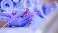
Free radicals have been implicated in many neonatal diseases. Historically, preterm infants have been exposed to high oxygen concentrations at birth in an effort to treat their immature lungs and surfactant deficiency. Endogenous antioxidant formation occurs primarily during the third trimester, leaving the preterm infant lacking defenses against oxidative stress. Disturbances in the balance of oxygen delivery and antioxidant availability have been implicated in chronic lung disease, retinopathy of prematurity, and intraventricular hemorrhage in preterm infants. Ongoing research seeks to determine optimal oxygen administration and treatment options to minimize these potentially devastating complications.
In the pediatric setting, evidence shows a link between free radical damage and neurologic outcomes in children with Down’s syndrome. Red blood cells in these children have an unbalanced antioxidant system, which may participate in certain neurologic manifestations. Nutritional supplements show promise in antioxidant regulation.
In adults, free radicals are implicated in aging, cardiovascular disease, cancer, and other diseases and disorders. The free radical theory of aging is based on what we currently know about lipid peroxidation and mDNA damage. The damage that occurs to many parts of the body during ischemia-reperfusion injury stems at least in part from free radical damage. Oxidative stress also is thought to trigger inflammatory bowel diseases, such as ulcerative colitis and Crohn’s disease.
In the intensive care setting, adults with respiratory distress, pancreatitis, and hyperglycemia have shown a higher incidence of oxidative stress. For example, fatigue associated with ventilator weaning has been associated with free radical damage, including lipid perioxidation and DNA and protein damage.
Implications for nursing practice
Free radicals are associated with many diseases and complications. But knowledge is power. Understanding free radicals at the cellular level can help us focus our care practices to potentially affect ROS production.
One significant nursing implication is recognizing that oxygen administration has risks. Oxygen is a drug and should be treated as such. Oxygen has proven in cerebral hypoxia, myocardial ischemia and infarction, impaired left ventricular function, wound healing, and decompression sickness. But its overuse and abuse can cause production of free radicals and subsequent damage. Thus, oxygen administration should be goal-directed and given as prescribed based on evidence-based objectives—and never without sufficient cause.
Also remember that physiologic stress is a factor involved in free radical production. As nurses, how can we alter our care practices to decrease patient stress? Can such practices as suctioning, positioning, and resuscitation affect ROS production? Research that would help answer these questions is limited—but this provides a great opportunity for nursing research.
Until evidence is available, we must use theory, knowledge, and critical thinking to guide our care for patients with potential imbalances of free radicals and antioxidants. Using the earlier examples of free radical damage, theory-based care can guide us as we minimize physiologic stress and free radical damage. For example, neonatal nurses can protect patients against oxygen poisoning by limiting exposure to high oxygen concentrations and diligently titrating oxygen delivery. Pediatric nurses can promote healthier outcomes in children with Down syndrome through effective nutritional education. Similarly, adult critical care nurses can take measures to ensure adequate perfusion for patients with ischemic injuries.
Ongoing research documents the benefits of antioxidant and scavenger therapies. As new medications, such as n-acetylcysteine, selenium, arginine, and other radical scavengers, are added to treatment regimens, nurses will need to become familiar with these therapies. Of course, all nurses can encourage, support, or participate in ongoing research on free radicals.
Jobeth Pilcher is a neonatal intensive care nurse at Baylor University Medical Center in Dallas, Texas and an adjunct online faculty member for several universities.
References
Amato M, Donati F. Update on perinatal hypoxic insult: mechanism, diagnosis and interventions. Eur J Paediatr Neurol. 2000;4(5):203-9.
Clark RH. Support of gas exchange in the delivery room and beyond: how do we avoid hurting the baby we seek to save? Clin Perinatol. 1999;26(3):669-81.
Davis KL, Martin E, Turko IV, Murad F. Novel effects of nitric oxide. Annu Rev Pharmacol Toxicol. 2001;41:203-36.
Dorweiler B, Pruefer D, Andrasi TB, Maksan SM, et al. Ischemia-reperfusion injury: pathophysiology and clinical implications. Eur J Trauma Emerg Surg. 2007;33(6):600-13.
Galloway JN, Aber JD, Erisman JW, Seitzinger SP et al. The nitrogen cascade. Bioscience. 2003;53(4):341-57.
Genestra M. Oxyl radicals, redox-sensitive signaling cascades and antioxidants. Cell Signal. 2007 Sep;19(9);1807-19.
Goodyear-Bruch C, Pierce JD. Oxidative stress in critically ill patients. Am J Crit Care. 2002 Nov;11(6):543-53.
Lawler JM, Cline CC, Hu Z, Coast JR. Effect of oxidative stress and acidosis on diaphragm contractile function. Am J Physiol. 1997;273(2 Pt 2):R630-6.
Majno G, Joris I. Cells, Tissues, and Disease Principles of General Pathology. (2nd ed.). Oxford University Press; 2004.
Poon HF, Calabrese V, Scapagnini G, Butterfield DA. Free radicals: key to brain aging and heme osygenase as a cellular response to oxidative stress. J Gerontol A Biol Sci Med Sci. 2004 May;59(5):478-93.
Sanz A, Pamplona R, Barja G. Is the mitochondrial free radical theory of aging intact? Antiox Redox Signal. 2006 Mar-Apr;8(3-4):582-99.
Smigielska-Kuzia J, Sobaniec W, Kulak W, Zawada B, et al. Antioxidant enzymes and lipid peroxides in children with Down syndrome. J Ped Neurol. 2007;5(2):117-20.
Tin W, Gupta S. Optimal oxygen therapy in preterm babies. Arch Dis Child Fetal Neonatal Ed. 2007;92:F143-F147. DOI:10.1136/adc.2005.092726.
Tortora GJ, Derrickson B. (eds.). Principles of Anatomy and Physiology. (11th ed.). New York: John Wiley; 2007.
Editor’s note: Figures from “Free Radicals”, published by the Neonatal Network, Volume 21, Number 7, November/December 2002. Reproduced with permission.


3 Comments.
Thank you for this information I now know what is free radicals and its implications in nursing care. We need more research in this.
well explained, thank you.
It cleared all my doubts, Excellent feature