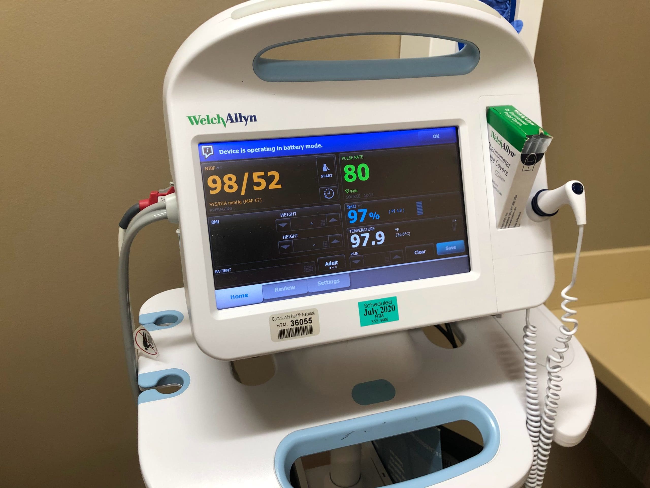
Anita Jenkins, age 54, arrives at the emergency department (ED) complaining of shortness of breath and chest tightness. An electrocardiogram (ECG) recorded within 6 minutes of her arrival shows normal ST segments; in the meantime, she reports that her chest discomfort has subsided. She’s started on supplemental oxygen at 4 L/minute and connected to a cardiac monitor; blood is drawn for cardiac biomarker testing.
Approximately 45 minutes later, the ST-segment alarm on her cardiac monitor rings, and the nurse detects ST-segment elevation in the anterior leads (V2, V3, and V4). Mrs. Jenkins says she’s short of breath and her chest feels tight again. A repeat 12-lead ECG confirms anterior-wall ischemia. Within minutes, she’s taken to the catheterization lab for a percutaneous coronary intervention (PCI).
Giving silent ischemia a voice
Without continuous ST-segment monitoring, the silent ischemia suffered by Mrs. Jenkins and other patients with acute coronary syndrome (ACS) might easily go undetected until irreversible myocardial damage occurs. About 80% to 90% of ECG-detected ischemic episodes are clinically silent. By revealing changes in the ST segment (the component that immediately follows the QRS complex on the ECG), continuous ST-segment monitoring can reveal both the severity and duration of ischemia, giving early warning of ischemic events.
The standard 12-lead ECG captures only a small snapshot (approximately 10 seconds) of the myocardial electrical cycle—a period too brief to show the waxing and waning of ST segments. Continuous ST-segment monitoring, in contrast, reveals ischemic events over time, providing a more accurate and complete picture of the patient’s myocardial physiology.
In 2004, the American Heart Association (AHA) published a standard that provides guidelines for continuous ST-segment monitoring to help improve outcomes for patients with ischemia. The information in this article reflects that standard.
Which patients benefit?
The best candidates for continuous ST-segment monitoring are patients with ACS who have unstable angina or acute myocardial infarction (AMI) with or without ST-segment elevation. Detecting myocardial ischemia early allows the healthcare team to reverse ischemia and thus prevent or interrupt myocardial cell death. In the ED, these patients initially may have a nondiagnostic ECG, but continuous ST-segment monitoring shows that a certain percentage have ST-segment changes within minutes to hours.
Continuous ST-segment monitoring also contributes to improved survival rates in AMI patients. Early identification of ischemic events gives caregivers the opportunity to initiate early reperfusion and take other actions to help keep the involved artery patent. For instance, it helps them assess patency of the infarct-related vessel after thrombolytic therapy and detects recurrent ischemic episodes after a PCI or infarct extension.
Postoperative monitoring
In patients who’ve undergone cardiac surgery, continuous ST-segment monitoring yields information about graft patency and the occurrence of perioperative MI. Generalized ST-segment deviation across several leads rather than isolated to a group of leads suggests postoperative pericarditis.
Other uses
Continuous ST-segment monitoring may reveal the extent of tissue injury in patients undergoing transmyocardial laser revascularization.
It also may benefit patients experiencing coronary vasospasm or ischemia related to hypoxia, as well as those recovering in the intensive care unit after cardiac surgery and other procedures.
Other uses of continuous ST-segment monitoring include:
• detecting perioperative ischemia in patients with a cardiac history who are undergoing other surgical procedures
• detecting abrupt closure and reocclusion of the affected vessel after PCI
• helping to differentiate cardiac and noncardiac chest pain
• helping predict failure to wean from mechanical ventilation.
Electrode placement
Consistent placement of electrodes and leads is crucial for accurate monitoring, because improperly placed electrodes can contribute to false alarms.
To help ensure accurate monitoring, take these steps when placing electrodes:
• At each electrode site, clean the patient’s skin with soap and water, rubbing the area slightly with a washcloth or gauze pad. You may use alcohol if the skin is oily, but be aware that alcohol can dry the skin and increase electrical resistance.
• Plot out the appropriate electrode position for each V lead.
• Make sure electrodes have moist gel inside.
Which leads to monitor
Monitoring all 12 leads provides more sensitive ST-segment monitoring. Research has shown that certain leads are more likely to reveal ischemic events associated with coronary artery occlusion. Here are some examples:
• To detect right coronary artery occlusion, the best lead to monitor is III, followed by aVF and II.
• Lead V2 or V3 is best for monitoring the left anterior descending coronary artery; lead V4 is less sensitive for this artery.
• Lead V1 or V2 is best for detecting reciprocal changes in posterior-wall ischemia. No specific lead is sensitive in detecting left circumflex coronary artery occlusion.
If a 12-lead monitoring system isn’t available, use leads II and V3 in a two-lead system and leads III, V3, and V5 in a three-lead system.
Detecting ST-segment changes
The ST-segment analysis point is 60 ms (0.06 sec) beyond the J point—located where the QRS complex ends and the ST segment begins.
To detect ST-segment changes effectively, be sure to monitor from the patient’s baseline (the point at which the ST-segment monitoring system is activated). If you have trouble determining whether the ST segment is above or below the baseline, try to identify the PR segment.
Be aware that body position can affect monitoring. Supine positioning is recommended, with the head of the bed elevated less than 45 degrees. If the alarm sounds, return the patient to the supine position and reassess before concluding that the alarm signals an ischemic event.
Always customize monitoring alarms to each patient. Preferably, set alarms with a 1-mm to 2-mm deviation above or below the baseline ST segment.
ST-segment “fingerprint”
Each patient has a unique pattern of ST-segment elevation or depression, called an ST-segment “fingerprint.”
• For patients experiencing an AMI, the lead that shows the maximum peak ST-segment elevation determines the fingerprint.
• For patients undergoing PCI, the single lead with peak ST elevation at the time of balloon catheter inflation determines the fingerprint.
Practice implications and recommendations
Incorporating continuous ST-segment monitoring into your clinical practice helps you determine if your patient is experiencing myocardial ischemia and aids implementation of appropriate interventions.
Healthcare providers and facilities can improve ST-segment monitoring practices by:
• purchasing user-friendly ECG equipment
• providing ongoing staff education on proper electrode placement, alarm setting, and ST-segment interpretation
• educating all nurses and physicians about the value of continuous ST-segment monitoring and the recent AHA practice standard
• developing protocols to specify the nurse’s response to ST elevation or depression (such as obtaining a 12-lead ECG to confirm validity of the deviation)
• reviewing applicable policies and procedures to develop proficiency standards
• undertaking audits to evaluate ST-segment monitoring practices.
Although the practice standard for continuous ST-segment monitoring specifically targets ED patients with ACS, it applies to all cardiac patients. By following this standard, you can help improve patient outcomes and contribute to a safer clinical environment.
Selected references
American Association of Critical-Care Nurses. Practice Alert: ST-segment monitoring. Available at: www.aacn.org/AACN/practiceAlert.nsf. Accessed December 19, 2006.
Drew BJ, Califf RM, Funk M, et al. Practice standards for electrocardiographic monitoring in hospital settings. Circulation. 2004;110:2721-2746.
Drew BJ, Funk M. Practice standards for ECG monitoring in hospital settings: executive summary and guide for implementation. Crit Care Nurs Clin North Am. 2006;18:157-168.
Drew BJ, Krucoff MW. Multilead ST-segment monitoring in patients with acute coronary syndromes: a consensus statement for healthcare professionals. Am J Crit Care. 1999;8(6):372-388.
Flanders S. Continuous ST-segment monitoring: raising the bar. Crit Care Nurs Clin North Am. 2006;18:169-177.
For a complete list of selected references, see February 2007 references.
Barbara (“Bobbi”) Leeper, MN, RN, CCRN, is a Clinical Nurse Specialist for Cardiovascular Services at Baylor University Medical Center in Dallas, Tex.