
You’ve been a registered nurse for 26 years and cared for dozens of breast cancer patients. You’ve listened as they poured out their feelings and fears. You’ve consoled them and prepared them for surgery. You’ve told them what to expect during chemotherapy and radiation and helped them cope with body-image changes and hair loss.
Suddenly, the tables are turned. You have no family history of breast cancer and no other risk factors, and have had yearly screenings. Yet now, at age 54, you find a lump in your right breast; a mammogram and ultrasound confirm an abnormality.
You undergo a breast biopsy. The doctor calls with the results: breast cancer.
You feel like you might faint. You think, “This can’t be happening to me!” Although you’ve taught many patients about breast cancer and its treatment, your knowledge base seems to have evaporated. Yesterday you could answer all your patients’ questions. Today, you have nothing but questions of your own.
For you and anyone else newly diagnosed with breast cancer—and for everyone who cares for them—this article promotes a better understanding of the disease. It gives a general overview of breast cancer screening, diagnosis, and treatment, highlighting information about new diagnostic and therapeutic developments you might not be familiar with.
Breast cancer basics
Breast cancer is the most common cancer in women and the second leading cause of cancer deaths. According to the American Cancer Society (ACS), about one in eight women will be diagnosed with breast cancer during their lifetime. Breast cancer also strikes men but in much lower numbers (1 in 100).
Breast cancer occurs in two basic types—noninvasive and invasive.
- The noninvasive type remains in the breast ducts or lobules, and is called ductal or lobular carcinoma in situ (DCIS or LCIS).
- Invasive cancer occurs when cancer cells spread, or metastasize, beyond the lining of the duct or lobule. Metastasis sites include the lymph nodes, bone, liver, lungs, and brain.
Usually, the cause of breast cancer is unknown. Occasionally, the disease clusters in families.
Risk factors
The risk of breast cancer increases with age. About 77% of women with breast cancer are older than age 50 at diagnosis. Persons at high risk include those who carry or develop certain genetic mutations and those with a family history of breast cancer. (See Breast cancer risk factors by clicking on the PDF icon above.)
The Breast Cancer Risk Assessment Tool can be used to calculate both a woman’s risk of developing invasive breast cancer over a 5-year period and her lifetime probability of developing breast cancer. Validated by several studies, it considers six factors—current age, age at menarche, previous breast biopsies, age at first live birth, history of breast cancer in first-degree relatives, and race or ethnicity. The tool is available at www.cancer.gov/bcrisktool.
Genetic risk factors
Breast cancer has a genetic basis in about 5% to 10% of cases, with multiple genetic defects implicated. Genes associated with an increased risk of breast cancer include:
- breast cancer 1, early onset (BRCA1)
- breast cancer 2, early onset (BRCA2)
- CHK2 checkpoint homolog (CHEK2)
- tumor protein p53 (TP53).
BRCA1 and BRCA2 are major genes whose mutations are linked to breast cancer. Normally, these genes function as tumor suppressor genes; if they mutate and allow unregulated cell growth, breast cancer may occur. About 36% to 85% of women with an altered BRCA1 or BRCA2 gene develop breast cancer. Blood testing can reveal BRCA1 and BRCA2 mutations and is commonly done in conjunction with genetic counseling.
The most common genetic mutation in cancer cells involves the TP53 gene. Normally, the p53 gene product recognizes damaged DNA and directs the cell to perform apoptosis (suicide). A p53 mutation leads to uncontrolled cell growth and subsequent cancer. More research is needed to find out if p53 testing should be recommended to help determine prognosis in breast cancer patients.
The risk of genetically based breast cancer is greatest in persons of Eastern-European Jewish background and in families where:
- multiple breast cancer cases have occurred
- members have been diagnosed with both breast and ovarian cancer
- members have been diagnosed with breast cancer at an early age
- one or more members have had two primary breast cancers
- males have had breast cancer.
Reducing breast cancer risk
Strategies to help detect and prevent breast cancer include regular screening for all women, lifestyle changes (such as weight control and exercise), preventive surgery (such as prophylactic mastectomy or oophorectomy), and chemoprevention with tamoxifen or raloxifene for certain women.
The STAR (Study of Tamoxifen and Raloxifene) trial, a breast cancer prevention trial, found that raloxifene (currently used to prevent and treat osteoporosis in postmenopausal women) works as well as tamoxifen in reducing breast cancer risk in high-risk postmenopausal women. Both drugs reduced invasive breast cancer risk by about 50%, but women receiving raloxifene had 36% fewer uterine cancers and 29% fewer blood clots than those receiving tamoxifen. Raloxifene is under review by the Food and Drug Administration (FDA) for approval as breast cancer chemoprevention.
Breast cancer screening
The ACS recommends the following screening guidelines for breast cancer:
- yearly mammograms starting at age 40
- clinical breast exams every 3 years for women in their 20s and 30s, and yearly for women ages 40 and older
- magnetic resonance imaging (MRI) scanning and mammography every year for women at higher risk (greater than 20% lifetime risk), starting at age 30. Women at moderately increased risk should talk with their primary care providers about the benefits and limitations of adding MRI screening to their yearly mammogram. (Yearly MRI screening isn’t recommended for women with a low lifetime breast cancer risk.)
In studies, MRI found breast cancer in women at an earlier stage (stage 0 to I) than in women who didn’t undergo MRIs (stage I to II). Overall MRI sensitivity in high-risk patients ranges from 71% to 100%, compared to 16% to 40% for mammography sensitivity. However, MRI isn’t recommended for general screening because of high false-positive rates.
Starting in their 20s, breast self-exam is an option for women. The ACS advises women to become familiar with how their breasts feel normally and report any change promptly to their healthcare providers.
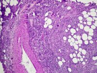
Assessment and diagnosis
Breast cancer may present as a palpable breast mass, breast pain, lymph node swelling, or skin changes, such as dimpling or redness. Evaluation of a breast abnormality usually starts with a clinical breast examination and mammography or ultrasonography, with MRI considered for some patients.
The next step is a needle biopsy or surgical excisional biopsy. To determine if the cancer has spread, the patient may undergo additional tests, such as X-ray, computed tomography (CT), a bone scan, and fluorodeoxyglucose (FDG) positron emission tomography (PET) integrated with CT (FDG-PET/CT).
Cancer staging
Cancer staging helps to determine appropriate treatment and estimate prognosis. Like other cancers, breast cancer is staged using the TNM system—tumor size (T), nodal involvement (N), and metastasis (M).
Once the TNM categories have been assigned, the clinician assigns an overall stage of 0, I, II, III, or IV. These stages identify tumor types that have a similar outlook and thus are treated in a similar way. For descriptions of breast cancer stages and treatments based on guidelines from the National Comprehensive Cancer Network (NCCN), see Breast cancer stage grouping.
Histopathologic type and grade
Other factors used to guide treatment and determine prognosis include the tumor’s histopathologic type and grade. Histopathologic breast cancer types include in situ, ductal, invasive, inflammatory, medullary, mucinous, papillary, lobular, and tubular.
Tumor grade refers to the extent to which the cancer cell resembles a normal cell. Breast cancer cells have three grades—low, intermediate, and high. In low-grade cancer, cells most resemble a normal cell and prognosis is more favorable. In high-grade cancer, cells look least like a normal cell and the prognosis is less favorable.
Hormone-receptor status
The tumor’s hormone-receptor status also influences prognosis and guides treatment. Testing can measure the number of estrogen and progesterone receptors in the tumor, which is reported as 0, 1+, 2+, or 3+. A value of 3+ is considered highly hormone-receptor positive and suggests the patient is more likely to respond well to antiestrogen therapy, such as tamoxifen or an aromatase inhibitor (AI). A value of 1+ means the tumor is borderline estrogen sensitive. A value of 0 predicts a poor response to antiestrogen therapy.
HER2 overexpression
A protein on the surface of all normal cells, human epidermal growth factor receptor-2 (HER2) helps regulate cell growth. About 20% of breast cancers overexpress HER2, making them more invasive, more resistant to chemotherapy, and more likely to recur. Over the past few years, determining the patient’s HER2 status has become important in managing breast cancer and guiding treatment.
One test to determine HER2 status measures HER2 using immunohistochemistry; the other test counts HER2 gene copies using a method called fluorescence in-situ hybridization. Such tests help predict if the patient would benefit from trastuzumab (Herceptin) or lapatinib (Tykerb) therapy.
Tumor markers
To help assess breast cancer, the patient may undergo testing for tumor markers, such as BCA (CA15-3), CA27.29, and CEA. Measuring these markers may aid diagnosis, help predict therapeutic efficacy, and detect cancer recurrence. High levels may indicate advanced or metastatic cancer.
Treatment
Breast cancer treatment options are constantly evolving. Treatment decisions may hinge on many factors, including the patient’s tolerance for the proposed therapy, patient preference, and comorbidities. To guide treatment and manage adverse effects, clinicians may use guidelines from the NCCN, American Society of Clinical Oncology, and Oncology Nursing Society. Clinical trials (listed at www.cancer.gov/clinicaltrials) may offer additional treatment opportunities.
Recently, treatment has become more individualized, based on specific features of the patient’s tumor. For instance, statistics show that as a group, patients with early-stage breast cancer don’t benefit from chemotherapy. Yet some of these patients have tumor characteristics that suggest chemotherapy would be helpful.
Multigene assays can help determine if breast cancer will respond to treatment. The MammoPrint test is used in patients younger than age 55 with stage I invasive cancer or stage II node-negative invasive cancer. Another assay, OncotypeDX, identifies a 21-gene cohort and is used for newly diagnosed stage I or II node-negative, estrogen-receptor-positive cancer. A low score means the patient may not benefit from chemotherapy; a high score suggests chemotherapy might be worthwhile.
Surgery and adjuvant therapy
Surgery is the definitive treatment for breast cancer. Usually, the patient receives adjuvant therapy in addition to the first therapeutic modality she undergoes. When surgery is the primary treatment, adjuvant therapy may consist of chemotherapy, radiation therapy, or antiestrogen therapy given afterward.
In neoadjuvant treatment, chemotherapy is given first to shrink a large tumor before surgery and thus allow a breast conservation technique (such as lumpectomy). It also may be used if surgery isn’t feasible at the time of diagnosis.
Breast reconstruction is an option for some women who’ve had mastectomies. Reconstruction may involve breast expanders, implants, autologous tissue reconstruction, or a combination.
Lymph node biopsy
Traditionally, patients with invasive breast cancer have undergone axillary lymph-node dissection to find out if cancer cells have spread to the axillary nodes. During this procedure, the surgeon removes the lymph node tissue that drains from the breast, and a pathologist determines if any of the nodes contains cancer. Full axillary node dissection can lead to such complications as lymphedema, neuropathy, and increased discomfort.
Sentinel lymph-node mapping offers an alternative to traditional axillary lymph-node dissection in patients with presumed early-stage disease, and can eliminate the need for full node dissection. This mapping technique identifies the sentinel (first) axillary lymph nodes (the ones most likely to be cancerous) based on the breast’s primary lymphatic drainage pattern. To map the sentinel nodes, a radioactive tracer and blue dye are injected in the breast tissue and carried by lymph fluid as it drains to the nodes. The nodes that take up the tracer and dye are identified and removed. A positive sentinel node indicates the need for a full axillary lymph-node dissection; negative sentinel nodes suggest other axillary nodes are cancer free.
In July, the FDA approved the GeneSearch BLN Assay—the first molecular-based laboratory test that determines if breast cancer has spread to surrounding lymph nodes. The test detects molecules that abound in breast tissue but are scarce in normal lymph nodes. Results are available in about 35 to 40 minutes while the patient is still on the operating table, instead of the 2-day wait and follow-up surgery now required. In a clinical trial, the GeneSearch BLN Assay accurately predicted that breast cancer had spread nearly 88% of the time in women with metastasis, and identified patients without metastasis 94% of the time.
Radiation therapy
Partial-breast radiation may involve internal, intraoperative, or external beam radiation.
- In internal partial-breast radiation (also called brachytherapy), radioactive seeds are inserted into the tumor bed.
- Intraoperative partial-breast radiation uses a linear accelerator to administer radiation to the tumor bed and surrounding area during surgery, after tumor removal.
- External beam partial-breast radiation begins with careful selection of the treatment field, followed by radiation delivered to the marked area with a linear accelerator.
Adverse effects of radiation therapy include fatigue and local skin reactions, such as discoloration, itching, soreness, and peeling. Late and long-term adverse effects also may occur. (See Late and long-term adverse effects of breast cancer treatments by clicking on the PDF icon above.)
Clinical trials are underway to compare partial-breast radiation to whole-breast radiation (the current standard). The latter is given by external beam to the entire breast and chest wall for 5 days weekly for up to 7 weeks. Partial-breast radiation is given in higher daily doses to the surgical cavity and margin over a shorter period. The trials may determine if the partial-breast technique is as effective as whole-breast radiation while speeding treatment time and causing fewer adverse effects.
Chemotherapy
Chemotherapy may involve a single antineoplastic drug or a combination of agents. Most patients with breast cancer receive chemotherapy in 2- to 3-week cycles, depending on the regimen. Concomitant administration of red and white blood cell growth factors may allow dose-dense chemotherapy, in which cycles are given closer together (such as every 2 weeks). Typically, breast cancer chemotherapy is given I.V., but some newer drugs used to treat metastatic disease are given orally.
Although chemotherapy targets cancer cells, it can also damage normal cells—especially the rapidly dividing cells of the GI tract, skin, hair, and bone marrow. Damage to normal cells can cause nausea, vomiting, diarrhea, hair loss, decreased blood counts, fatigue, and neuropathy.
New techniques have been developed to administer chemotherapy. For instance, using nanotechnology, one drug maker manufactures paclitaxel in an albumin-bound form. The FDA has approved this drug form for metastatic breast cancer treatment. Called paclitaxel protein-bound (Abraxane), it’s the first in the new category of protein-bound particle drugs.
Antiestrogen therapy
Patients with estrogen- or progesterone-positive breast cancer may be candidates for antiestrogen therapy, which reduces the amount of estrogen available to cancer cells. One type of antiestrogen therapy uses selective estrogen-receptor modulators, such as tamoxifen. By blocking estrogen receptors in breast cancer cells, tamoxifen stops estrogen from binding to these receptors and stimulating cancer cell growth. Tamoxifen can be given to premenopausal or postmenopausal women. Adverse effects may include hot flashes, coagulopathies (such as deep vein thrombosis), and endometrial cancer.
In the late 1990s, antiestrogen therapy involving AIs became available for postmenopausal women with estrogen- or progesterone-positive breast cancer. Before menopause, the ovaries produce most of the body’s estrogen; after menopause, estrogen comes from androgen’s conversion to estrogen, with the enzyme aromatase regulating a step in this process. AIs (which include anastrozole, letrozole, and exemestane) block aromatase function, halting estrogen production. Adverse effects include hot flashes, arthralgia, myalgia, and osteoporosis.
AIs have begun to replace tamoxifen as standard treatment for estrogen- or progesterone-positive breast cancer. The ATAC (Arimidex, Tamoxifen, Alone or in Combination) trial and other large clinical trials found AIs more effective than tamoxifen in postmenopausal patients with estrogen-receptor-positive cancer.
Targeted therapies
Targeted therapies use agents that interfere with specific molecules involved in carcinogenesis and tumor growth. By focusing on cancer-specific molecular and cellular changes, these therapies may be more effective than current treatments and may cause less harm to normal cells. Recent advances in targeted therapy for breast cancer include the drugs trastuzumab, bevacizumab, and lapatinib.
Trastuzumab (Herceptin) targets HER2-positive cancer cells. By binding to the HER2 receptor on the cell surface, it may trigger the body’s defense system to destroy that cell. The drug interrupts the cell’s growth signal, halting cell proliferation. Adverse effects include flulike symptoms, nausea, and cardiac changes that can lead to heart failure.
Bevacizumab (Avastin) stops tumor growth by targeting and inhibiting the function of vascular endothelial growth factor, a protein that promotes angiogenesis. In combination with paclitaxel, bevacizumab is used for metastatic breast cancer. Studies show the combination improves progression-free survival. Possible adverse effects of bevacizumab include bleeding, hypertension, proteinuria, weakness, pain, and fatigue. Lapatinib (Tykerb) is approved in combination with capecitabine (Xeloda) to treat HER2-positive metastatic breast cancers that don’t respond to chemotherapy or trastuzumab. Lapatinib binds to epidermal growth factor receptor-1 and HER2 receptors, stopping the cell from growing. The combination of lapatinib and capecitabine delayed breast cancer progression for nearly twice as long as capecitabine alone (4.4 months vs. 8.4 months). Possible adverse effects of the drug combination include diarrhea, nausea, vomiting, rash, and hand-foot syndrome.
Surviving breast cancer
Hearing that you or a loved one has breast cancer is a shock. But breast cancer usually doesn’t equate to death. Earlier detection through screening, increased awareness, and improved treatment have led to a continuing decline in mortality. According to the ACS, 5-year survival for women based on breast cancer stage at diagnosis is:
- 88.5% for all cancers
- 98.1% for localized cancer (stages I and II)
- 83.1% for cancer with regional spread (stage III)
- 26.0% for cancer with distant spread (stage IV).
More than 2 million people in the United States are breast cancer survivors, and the number continues to grow. Urge all patients who’ve had breast cancer to get routine follow-up care. Such care allows early detection of a recurrence or a new primary cancer and helps ensure that the patient is assessed and treated for late and long-term effects of cancer treatment.
Survivors know a diagnosis of breast cancer is a lifelong journey. Uncertain of the future, most live with the fear that cancer will recur. As nurses, we have the chance to support them from the time of diagnosis through treatment—and beyond.
Selected references
American Cancer Society. Breast cancer facts & figures, 2005-2006. Available at: www.cancer.org/downloads/STT/CAFF2005BrF.pdf. Accessed August 22, 2007.
American Cancer Society. What are the risk factors for breast cancer? Availableat:www.cancer.org/docroot/CRI/content/CRI_2_4_2X_What_are_the_risk_factors_for_breast_cancer_5.asp?sitearea=. Accessed August 22, 2007.
Muss H. Targeted therapy for metastatic breast cancer. N Engl J Med. 2006;355(26):2783-2785. National Cancer Institute. Breast cancer (PDQ®): treatment. Available at:cancer.gov/cancertopics/pdq/treatment/breast/HealthProfessional. Accessed August 22, 2007.
Ravdin P, Cronin K, Howlader N, et al. The decrease in breast-cancer incidence in 2003 in the United States. N Engl J Med. 2007;356(16):1670-1674.
Saslow D, Boetes C, Burke W, et al, for the American Cancer Society Breast Cancer Advisory Group. American Cancer Society guidelines for breast screening with MRI as an adjunct to mammography. CA Cancer J Clin. 2007;57(2):75-89.
For a complete list of selected references and to learn more about treatment of early-stage breast cancer, visit www.AmericanNurseToday.com.
The authors are Oncology Nurse Practitioners at the Marshfield Clinic, which has centers throughout Wisconsin. Julene Diedrich, MSN, RN, FNP, AOCNP, and Jessica Engel, MSN, RN, FNP, AOCN, work at the Marshfield center. Jill Depke, MSN, RN, ANP, AOCNP, works at the Weston center. The authors do not have any financial arrangements or affiliations with any corporations offering financial support or educational grants for continuing nursing education activities.


2 Comments.
Felicitaciones ala colega que nos recuerda una de las funciones propias de la enfermeria, me gustaria contactarla para decirle que en Colombia estamos interesados en investigar en este tema.
Thanks for reminding all of us how important it is to keep up with breast health. I have an acquaintance who is living proof that SELF exams really do work, too! Excellent article