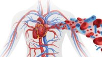
Two hours into your shift in the emergency department (ED) at a level-one trauma center, the medical communication radio sounds an alarm: Medics are on their way in with a 31-year-old male construction worker who was hit by a bulldozer and suffered a crushed pelvis. In the field, the patient’s pulse is 135 beats/minute, blood pressure 82/44 mm Hg, and oxygen saturation 99%. He is intubated and being ventilated manually, with two large-bore I.V. lines infusing lactated Ringer’s solution.
Some experts predict trauma will be the second leading cause of death by 2020. Hemorrhage is the leading cause of early preventable deaths from trauma, accounting for 50% of deaths in the first 6 hours after injury.
Finding and stopping the bleeding is the cornerstone to treating bleeding trauma. For the patient in our scenario, this might involve “binding” the pelvis with a commercial device or sheet to tamponade venous bleeding, along with angiography to embolize arterial bleeders. While the team tries to stop the bleeding, what’s the best way to resuscitate this patient? Historically, shock from blood loss has been treated with aggressive volume resuscitation using crystalloids and blood transfusions. Coagulopathy was thought to occur late in the course of trauma resuscitation, resulting from clotting factor consumption.
But a 2003 study described another mechanism for trauma-associated coagulopathy—acute traumatic coagulopathy (ATC). This condition results from low-flow shock states related to blood loss that activates anticoagulant and fibrinolytic pathways. A response to hemorrhagic shock, ATC may arise as a way to prevent vascular bed thrombosis; it can lead to systemic anticoagulation. ATC is exacerbated further by dilution resulting from fluid resuscitation (clotting-factor depletion), hypothermia (which impairs thrombin production and platelet function), and acidosis (which hinders thrombin production).
ATC occurs early after injury; patients presenting with ATC are eight times more likely to die during the first 24 hours after admission than patients who don’t have coagulopathy on arrival. The 2003 study found that 25% of injured patients were coagulopathic on ED arrival before receiving I.V. fluids.
Damage-control resuscitation
Along with a growing understanding of ATC, experience with combat casualties during the Iraq and Afghanistan conflicts has prompted a challenge to traditional trauma resuscitation methods. Resuscitation practice is starting to emphasize early blood-component therapy, including massive transfusion (MT), along with tolerance of moderate hypotension until bleeding is controlled. Called damage-control resuscitation (DCR), this strategy was proposed in 2005 at the U.S Army’s Institute of Research Conference.
DCR combines rapid control of bleeding, minimal crystalloid administration, permissive hypotension, and early hemostatic resuscitation with blood components that approximate whole blood. The goals of DCR are to:
- achieve definitive hemostasis
- aggressively correct hemorrhagic shock and ATC
- avoid iatrogenic injury.
Understanding permissive hypotension and hemostatic resuscitation
Permissive hypotension, which precedes surgical control of bleeding, allows systolic pressure to stay below normal (80 to 100 mm Hg) by limiting crystalloid fluid resuscitation. It’s based on the theory that raising blood pressure before the forming thrombus stabilizes may dislodge the thombus and cause rebleeding.
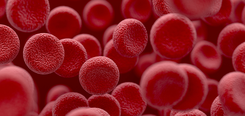
Hemostatic resuscitation refers to immediate or early administration of balanced blood components to severely injured patients in ratios approaching those of whole blood. A commonly used ratio is 1:1:1, meaning 1 unit of packed red blood cells (PRBCs) to 1 unit of plasma to 1 unit of platelets. For early ATC management, blood products take priority over crystalloid resuscitation.
Massive transfusion
MT is the immediate transfusion of blood and coagulation components in amounts physiologically similar to those in whole blood transfusions. MT promotes early aggressive administration of predefined blood components before laboratory tests suggest blood loss or confirm coagulopathy. MT initiation is based on the trauma mechanism, patient’s injuries, physiologic data, or a combination.
While bleeding is being controlled surgically, minimizing traumatic coagulopathy with MT is a key resuscitation goal for patients with traumatic hemorrhagic shock. Traditionally, MT has been defined as 10 units of PRBCs (equivalent to the blood volume of an average-size adult) transfused over 24 hours. But an updated definition of MT is 10 units of PRBCs given during the first 6 hours after ED admission. (About 80% of patients who die from hemorrhage do so during this critical period.) To keep up with ATC, blood volume initially lost from injury and ongoing blood loss must be replaced aggressively. Starting hemostatic MT late (trying to catch up) is linked to decreased effectiveness, increased overall product use, and higher mortality.
Recent U.S. military and observational multicenter civilian studies show improved outcomes with high blood product ratios—commonly 1 unit of plasma to 1 unit of PRBCs during MT. A plasma-to-PRBC ratio less than 1:1 may worsen existing coagulopathy by further diluting coagulation factors. All PRBCs and plasma given during MT should be transfused as fast as possible through a warming device.
Platelets are given in ratios of 1:1, or 2 units of PRBCs to 1 unit of platelets, where one bag of apheresis (single-donor) platelets equals 5 to 8 units.
Early vigorous transfusion of high ratios of blood and coagulation factors given up front may
decrease total transfusion requirements, so less blood may be needed overall. This protects the patient from unnecessary and potentially dangerous blood exposure and saves a limited resource. (See Complications of massive transfusion by clicking the PDF icon above.) Patients who don’t need traditional MT but still have substantial bleeding (defined in one study as the need for more than 4 units of PRBCs within 4 hours of ED admission) also may benefit from early high product ratios.
MT protocols
Management of MT with a specific protocol may lower patient mortality. Using an MT protocol (MTP) promotes more effective MT, improving blood-administration timing by optimizing systems flow to achieve rapid blood-product availability. MTPs support high component ratios, enhance communication to minimize blood-product delivery delays, and optimize outcomes by accomplishing earlier transfusion. MTPs also include instructions for administering cryoprecipitate, activated factor 7, or tranexamic acid (an antifibrinolytic that prevents clot breakdown). In one study, MTPs improved mean time to blood-product availability so effectively that deaths decreased from 45% to 19%.
MTPs were created to expedite empiric transfusion of PRBCs and coagulation factors without the need to wait for laboratory results. At many hospitals, traditional coagulation tests may take 1 hour, which is too long to guide MT effectively.
MTP activation
An attending trauma, emergency, or anesthesia physician activates the MTP—ideally, as soon as possible after ED admission. Research is ongoing to pinpoint the most accurate and readily available triggers for activation, including scoring systems. (See Triggers for initiating massive transfusion by clicking the PDF icon above.)
Whether and when to activate the MTP for patients who aren’t obviously exsanguinating isn’t always straightforward. Patients with normal vital signs who have internal bleeding and are at risk for hemorrhage, ongoing coagulopathy, and shock may not be recognized immediately. However, overtriaging (overestimating trauma severity) is preferable to undertriaging (underestimating severity).
Once the MTP is activated, transfusion personnel immediately start to assemble blood products in predefined ratios, which are delivered to the patient location in a series of designated coolers as quickly as possible. When the first cooler arrives, staff check the contents and administer the products promptly without further orders, while transfusion personnel immediately start preparing the second cooler for delivery and administration. The same process occurs with subsequent coolers. Patients receive transfusions continuously per protocol until they’re hemodynamically stable and bleeding is controlled; no target hemoglobin value or other laboratory value is used. The MTP continues with ongoing blood-product delivery until the attending physician stops it.
Clinical scenario continued
When the patient with the crushed pelvis arrives at the trauma center, the team checks and secures his airway, breathing, and circulation according to the trauma primary survey. He remains tachycardic, hypotensive, pale, and diaphoretic. The team applies a pelvic binder to tamponade bleeding and consults an interventional radiologist for emergent angiographic embolization. Although the patient’s blood pressure is low at 90/50 mm Hg, the trauma surgeon orders lactated Ringer’s solution from the field to be saline-locked, to decrease further bleeding and dilution of coagulation factors.
The trauma surgeon then activates the MTP. When the blood-product coolers arrive, you check each one and immediately begin transfusing the blood products with a rapid transfuser machine for speed and warming. Transfusions continue while the interventional radiologist embolizes three vessels to contain pelvic bleeding. As the embolization is completed, bleeding is controlled and the patient is hemodynamically stable (heart rate 88 beats/minute, blood pressure 119/75 mm Hg, and skin pink and dry). The trauma surgeon then ends the MT.
By this point, you’ve transfused 24 units of PRBCs, 24 units of plasma, and 4 units of apheresis platelets, for a transfusion ratio of 1 unit PRBCs to 1 unit plasma to 1 unit platelets. (One bag of apheresis platelets equals 5 to 8 units of platelets.) When the patient arrives at the intensive care unit, he is warm (36.8º C [98.2º F]) and without acidosis (pH 7.41, base deficit +1). His hemoglobin and
coagulation values are normal (hemoglobin 13 g/dL, platelets 151,000/µL, International Normalized Ratio 1.3). He continues to progress, and on hospital day 4, he is transferred to a medical unit.
Nursing roles and responsibilities
Nurses need to understand best practices for hemostatic resuscitation of trauma patients, because once the MTP is activated, nurses commonly manage the MT. Delayed or disorganized transfusion may lead to deficient product ratios, promoting a less favorable outcome. Early achievement of hemostasis using high blood-product ratios decreases overall blood-product requirements by addressing coagulopathy quickly. Once hemostasis is achieved, aggressive transfusion should be reevaluated.
Refinement of ideal trauma resuscitation strategy is ongoing. Hemostatic resuscitation with predefined ratios using MTPs may improve hemorrhagic shock outcomes. Multiple studies show decreased mortality when bleeding is controlled and patients receive high ratios of blood products within the first 6 hours of admission.
Click here for an example of an MTP, the typical contents and sequence of blood-product coolers, and a list of selected references.
The authors are Clinical Nurse IVs in the Crisis/RRT program at the Queen’s Medical Center in Honolulu, Hawaii. Christy Passion is also a Clinical Nurse IV in the surgical intensive care unit.