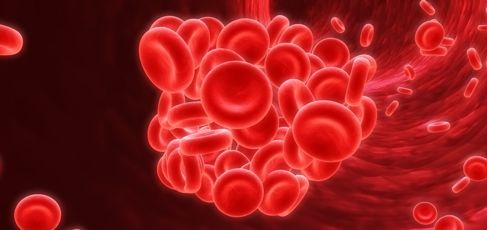
Pulmonary embolism (PE) occurs when a pulmonary artery becomes blocked—usually by a blood clot that has broken free from its site of origin and embolized or migrated to the lungs. If misdiagnosed, unrecognized, or untreated, PE can cause death quickly—within just an hour. It’s fatal in up to 26% of cases.
Massive PE, defined as causing 50% or more occlusion of the pulmonary capillary bed, can result in obstructive shock with systemic hypoperfusion (low cardiac output and acute pulmonary hypertension with right ventricular failure). It must be remedied immediately to save the patient’s life.
The true incidence of PE is unknown. Estimates range widely from 500,000 to 780,000 cases annually in the United States. Undiscovered PE is a frequent autopsy finding, with estimates of more than 400,000 undiagnosed postmortem cases. Because of its high mortality and the diagnostic challenges it poses, clinicians must be diligent in preventing PE, recognizing it early, and providing prompt and appropriate treatment.
Pathophysiology
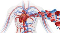
The usual underlying cause of PE is deep vein thrombosis (DVT) in a lower extremity, the pelvis, or even an upper extremity. (See How a pulmonary embolism evolves by clicking on the PDF icon above.)
DVT, in turn, typically results from one or more of these conditions: venous stasis, blood hypercoagulability (increased clotting), or endovascular damage. Known collectively as Virchow’s triad, these conditions can stem from a range of situations. For instance, venous stasis and hypercoagulability may result from immobilization or regional states of low blood flow caused by trauma (especially to the lower legs), burns, shock, obesity, or heart disease. Endovascular damage may stem from central venous access or venous procedures, when invasion and manipulation of the intimal wall of the vein increase platelet aggregation or fibrin production, causing clot formation. (See Risk factors for Virchow’s triad and PE by clicking on the PDF icon above.)
Complications
Pulmonary infarction may occur, although it usually arises later, during heparin infusion or other treatments. Presumably, infarction relates to initial clot instability and breakdown, resulting in further distal embolization that occludes the subpleural blood vessels. Pulmonary infarction manifests on chest X-ray as a pleural-based, wedge-shaped density called a Hampton hump. This density reflects parenchymal necrosis (peripheral regions where decreased blood supply has destroyed tissue) and results from pulmonary infarction to peripheral lung regions. Necrosis is associated with chest pain and hemoptysis. Life-threatening hemoptysis may occur from asphyxiation and respiratory arrest, not from the hemodynamic consequences of blood loss.
Signs and symptoms
Often, PE and DVT don’t cause symptoms, making these conditions challenging to detect. In many patients, even extensive testing fails to confirm these disorders.
When PE signs and symptoms do occur, they may be nonspecific, and include:
- shortness of breath
- dry cough
- hemoptysis
- diaphoresis or sudden chest pain that worsens with deep inspiration. The chest pain typically accompanies lung infarction, as the pleura is the only part of the lung that can sense pain.
More severe clinical findings may include hypotension, hypoxemia, and loss of consciousness. Classically, these occur with a massive PE, multiple PE, or in situ clot propagation.
DVT causes signs and symptoms in only about half of those affected. Signs and symptoms of DVT in the leg include leg or calf pain, especially on dorsiflexion (positive Homans’ sign); redness or discoloration; and increased warmth and swelling in the affected leg. Early DVT diagnosis and management are crucial, helping to prevent emboli from migrating to the pulmonary vasculature.
Diagnosis
Clinicians may use a scoring tool such as the Geneva score to aid PE diagnosis. The simplified Geneva score is based on nine patient risk factors and clinical variables; patients with a total score of two points or less are considered unlikely to have PE.
For instance, where each item carries one point, suppose a patient:
- has a history of a previous DVT or PE (1 point)
- is age 65 or older (1 point)
- has hemoptysis (1 point).
This patient’s total score is 3 points, signifying an elevated prediction of PE that warrants further diagnostic intervention.
Specific tests
In the hospital setting, the most common radiologic test is a chest X-ray. However, it’s neither sensitive nor specific for PE. More often, an X-ray helps identify conditions that may mimic PE. With PE, a chest X-ray usually shows minimal or no atelectasis. Rarely, more specific indicators of PE may appear on X-ray, such as the Westermark sign (reduced blood volume), hyperlucency (reduced density), and the Hampton hump (which appears in peripheral lung regions). Ultrasonography (including duplex ultrasonography) may be used to locate primary clots in extremities.
The deep pelvic veins are hard to evaluate with these noninvasive tests, so additional tests may be done if clinical suspicion of PE is high. Lung ventilation/perfusion (V/Q) scanning with radioisotopes may detect a blood clot that is impeding blood flow to the lung. Although useful when test results reveal a normal or high probability, an intermediate-probability V/Q result is inconclusive for PE.
Pulmonary angiography may be used to show where a clot is cutting off blood supply to a vessel. A contrast-enhanced helical computed tomography scan may be less invasive and now is more commonly used to visualize blood clots and diagnose PE.
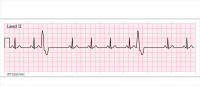
Additional tests, such as echocardiography, electrocardiography, magnetic resonance imaging, and laboratory tests (such as D-dimer testing), may be ordered to detect other conditions or to further rule out, support, or confirm PE.
Since its introduction in the 1990s, the D-dimer test has become important when thrombotic disorders are suspected, although positive results can be hard to interpret. A negative result virtually rules out thrombosis; a positive result may indicate thrombosis but doesn’t exclude other conditions, such as liver disease, renal or heart failure, or cancer. Therefore, a positive D-dimer test generally warrants additional testing to exclude or support other diseases and conditions.
Management
The goal of treatment is to halt PE evolution by reducing the propagation of an existing clot or preventing a new clot from forming and embolizing. A patient with confirmed PE may receive anticoagulants or thrombolytics or may undergo surgery.
Hemodynamically significant PE frequently is an indication for thrombolysis (clot lysis) rather than just anticoagulant therapy. In situ clot propagation can be treated effectively with anticoagulation if intervention begins early and therapeutic goals are achieved and maintained. Use of heparin nomograms may help clinicians reach these goals and minimize failure.
Although heparin and warfarin don’t lyse existing clots, they help prevent clot propagation and allow the body to lyse the clot naturally. The natural lytic process takes several months to break down the clot and recannulize the vessel, so prolonged anticoagulation is needed. Warfarin (Coumadin) is given orally but doesn’t take effect for several days; generally, it’s given in combination with I.V. heparin until an appropriate International Normalized Ratio (INR) is reached. The goal of anticoagulant therapy is to achieve an INR of 2 to 2.5 for a range of 3 to 6 months. Possible adverse effects of anticoagulants include bruising and bleeding.
The Joint Commission notes that anticoagulants are more likely than other medications to cause harm because of their complex dosing, insufficient monitoring, and inconsistent patient adherence. Regular INR monitoring and teaching patients about therapy are essential to helping them achieve desired outcomes.
Thrombolytics
Thrombolytics dissolve or lyse clots acutely but can cause immediate and severe bleeding. They’re reserved for hemodynamically unstable patients. Fast-acting thrombolytics—t-PA (alteplase) and r-PA (reteplase)—are the most common thrombolytics used.
IVC filter
An inferior vena cava (IVC) filter may be used to prevent a clot from embolizing to the lung. In this procedure, a catheter is used to position a filter in the IVC. Practice guidelines for retrievable IVCs include three basic indications for IVC placement: absolute, relative, and prophylactic.
- Absolute indications include recurrent acute or chronic venous thromboembolism and contraindications, complications, or inability to maintain appropriate anticoagulant therapy.
- Relative indications include a free-floating DVT, massive PE, ineffective or complications of anticoagulant therapy, and chronic PEs treated with thromboendarterectomy.
- Prophylactic indications include such conditions as trauma, prolonged surgical procedures, and certain medical conditions (for instance, atrial fibrillation). (See Case study: A fall, a fracture, and a sudden crisis by clicking on the PDF icon above.) Retrievable IVC filters should be removed as soon as protection from PE isn’t needed.
Embolectomy
Embolectomy (clot removal) may be done with a Fogarty catheter or may be achieved surgically through an incision in the vessel to remove the clot manually. Generally used as a last resort, the procedure may be required to remove a large clot or when thrombolytics are contraindicated or ineffective.
Standard practice for DVT prophylaxis
DVT prophylaxis should be implemented in all hospital patients, especially those with decreased mobility (such as those in critical care units or rehabilitation or extended-care facilities). Adequate hydration is vital to reducing blood viscosity and hypercoagulability. Early removal of central venous lines reduces the risk of endovascular damage. In immobilized patients, range-of-motion exercises and physical therapy (especially to the lower extremities) should begin as soon as possible to promote circulation and reduce venous stasis.
External pneumatic compression (EPC) devices, which deliver rapid inflation with intermittent pneumatic compression, may be used in patients confined to bed rest or unable to walk due to injury or illness. Numerous studies show the efficacy of EPC, with improvement of local hemodynamic flow the primary benefit. Also, EPC increases fibrinolytic activity in systemic blood.
Subcutaneous weight-based heparin dosing or routine administration of low-molecular-weight heparin (LMWH) has been shown to prevent DVT. However, thrombosis prophylaxis with heparin or LMWH augmented with EPC devices was found to be more effective in preventing DVT compared to LMWH alone. Trauma, integumentary disruption (as from burns), or external fixation devices or casting for bone stabilization may preclude the use of EPC, increasing the patient’s risk of DVT.
Nursing considerations
For any hospital patient, DVT prevention and risk reduction are essential nursing goals. Whenever possible, only minimal sedation, if any, should be used as a way to keep patients more active. Neuromuscular blocking agents should be avoided because they’ve been linked to DVT. Use of sedation scales may provide a more consistent approach to sedation dosing, as might sedation and analgesia protocols.
Implementing and maintaining prophylactic measures as a standard practice for preventing DVT are essential. Protocols for ventilator management and weaning also may be crucial to restoring activity levels and liberating patients from mechanical ventilation.
Make sure you’re familiar with DVT and PE risk factors and signs and symptoms. Be aware that when PE is confirmed or strongly suspected, treatment must begin immediately. Be sure to monitor patients for pain, anxiety, and anticoagulant side effects (such as bleeding), and provide appropriate interventions.
Selected references
American College of Chest Physicians. Antithrombotic and Thrombolytic Therapy, 8th Ed: ACCP Guidelines. Chest. June 1, 2008; (suppl 6):133. http://chestjournal.chestpubs.org/content/133/6_suppl.toc. Accessed August 4, 2010.
Habashi NM, Andrews PL, Scalea TM. Therapeutic aspects of fat embolism syndrome. Injury. 2006;37(suppl 4):S68-73. Review. Erratum in: Injury. 2007;38(10):1224.
Shaughnessy K. Massive pulmonary embolism. Crit Care Nurse. 2007;27(1):39-53.
Both authors work at R Adams Cowley Shock Trauma Center in Baltimore, Maryland. Penny L. Andrews is a staff nurse; Nader M. Habashi is medical director of the Multitrauma Critical Care unit. The planners and authors of this CNE activity have disclosed no relevant financial relationships with any commercial companies pertaining to this activity.


1 Comment.
This is agreat post. i actually like how you elaborated on this