Correctly identifying and interpreting cardiac rhythms can pose a challenge even for the most experienced nurses—let alone those who don’t read rhythm strips on a regular basis. Atrioventricular (AV) blocks can be particularly confusing and hard to interpret. But by gaining a basic understanding of the heart’s conduction system and electrocardiography, you can recognize these arrhythmias more easily.
Basic cardiac conduction and electrocardiography
In a normally functioning heart, an impulse initiated in the sinoatrial (SA) node takes 0.12 to 0.20 sec (120 to 200 msec) to travel through the atria, AV node, bundle of His, and bundle branches, from which the ventricles receive the impulse and begin depolarization (contraction). This duration (0.12 to 0.20 sec) is called the PR interval; on the electrocardiogram (ECG), it’s measured from the start of the P wave to the start of the QRS complex. Normally, the QRS complex is 0.06 to 0.11 sec (60 to 110 msec) wide. A QRS complex wider than 0.11 sec indicates either a bundle branch block or a QRS complex of ventricular origin.
To identify any arrhythmia, you need to understand and be able to recognize sinus rhythm, including its normal parameters. These parameters include:
- P waves occurring regularly at a rate of 60 to 100 per minute
- QRS complexes occurring regularly at a rate of 60 to 100 per minute
- 1:1 relationship between P waves and QRS complexes (for every P wave, a QRS complex is conducted)
- normal PR-interval duration (0.12 to 0.20 sec)
- normal QRS width (0.06 to 0.11 sec).
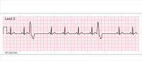
These normal parameters appear in the rhythm strip below. P waves and QRS complexes occur regularly at a rate of approximately 70 per minute. P waves and QRS complexes have a 1:1 relationship; the PR interval is approximately 0.14 sec and QRS width is approximately 0.08 sec.
What happens in AV block
AV block represents either delayed impulse transmission, an altered rate of impulse transmission, or complete blockage of impulse transmission from the atria to the ventricles. An altered rate or a blockage may result from an anatomic (physiologic or pathophysiologic) or functional (iatrogenic) impairment of the conduction system; it may be transient or permanent. The delay manifests in the PR interval and the relationship between P waves and QRS complexes. A wide variety of conditions can lead to AV block. (See Major causes of AV block by clicking the PDF icon above.)
During an AV block, the SA node generally functions properly. While impulse formation is normal, impulse conduction through the AV node, bundle of His, or bundle branches is abnormal. The PR interval and its relationship to the QRS complex deviate from normal, with the type of deviation indicating the type of AV block. Using sinus rhythm as a template, you can focus on these deviations when interpreting the type of AV block. (However, know that AV blocks also can occur with sinus bradycardia or sinus tachycardia.)
AV blocks are best interpreted by looking at the PR interval and relationship of P waves to QRS complexes. Do QRS complexes result from P waves (as expected), or are P waves present without QRS complexes?
Types of AV blocks
AV blocks occur in several types:
- first-degree
- second-degree type I
- second-degree type II
- variations of second-degree block
- third-degree.
Arrhythmias with AV block (except third-degree AV blocks) technically are called sinus rhythms with an X type of AV block—for example, sinus rhythm with a first-degree AV block. But in everyday communication and documentation, they’re simply referred to as first-degree AV block, second-degree AV block type I, and so on.
First-degree AV block
In first-degree AV block, impulse transmission from the atria to the ventricles slows slightly in the AV node. This causes a PR interval that exceeds the normal upper parameter of 0.20 sec. In essence, it’s simply a sinus rhythm with a PR interval longer than 0.20 sec (sometimes significantly longer), with all other parameters normal.
P waves and QRS complexes have a 1:1 relationship, as shown in the strip below.
Second-degree AV blocks
Two main types of second-degree AV blocks exist—Mobitz type I (frequently called Wenckebach) and Mobitz type II. Two additional variations (discussed later) also may occur.
In second-degree blocks, the part of the conduction system that progressively slows or blocks impulses (or does both) may be the AV node, bundle of His, or bundle branches. Because some impulses don’t reach the ventricles, these blocks always have missing QRS complexes, which causes an irregular-looking rhythm. On the ECG, look at the PR interval to help distinguish second-degree type I from second-degree type II blocks. P waves
occur consistently because the SA node is functioning properly.
Second-degree Mobitz type I
This block has three identifying features:
- progressive PR-interval lengthening before each conducted QRS complex, until a P wave finally occurs without a subsequent QRS complex
- grouping of QRS complexes due to the blocked or dropped impulse
- irregular occurrence of QRS complexes (an irregular rhythm) due to the blocked impulse, which causes a pause. Typically, QRS complexes are within normal width parameters, as shown in the strip below.
Second-degree Mobitz type II
This arrhythmia has four identifying features:
<ul
- P waves occurring consistently with some blocked impulses
- PR intervals of conducted QRS complexes that are of consistent length
- blocked impulses and missing QRS complexes
- QRS complexes occurring irregularly due to blocked impulses.
Intermittent and unpredictable blocking of impulses causes the missing QRS complexes. Parts of the conduction system that may be involved in this type of block are the AV node, bundle of His, and bundle branches. As a result, QRS complex widths may vary. If the blockage is in the AV node or bundle of His, QRS complexes have a normal width. If the block is in the bundle branches, typically one bundle branch is blocked all the time while the other bundle branch is blocked periodically. When this occurs, conducted QRS complexes are wide, with a typical bundle-branch-block appearance. This is the most common blockage mechanism for second-degree AV block type II. The rhythm strips below show this arrhythmia.
Variations of second-degree AV block
Two variations of second-degree AV blocks can occur:
- those with 2:1 conduction
- those with advanced-grade or high-grade block.
In blocks with 2:1 conduction, two P waves occur for every QRS complex, indicating every other impulse is blocked, as shown in the strip below. With every other P wave being conducted, this rhythm has regularly occurring QRS complexes. This variation can occur in either type I or type II AV block. However, because this arrhythmia lacks two conducted P waves in a row, types I and II can’t be differentiated, so it’s simply called second-degree AV block with 2:1 conduction.
In advanced-grade or high-grade second-degree AV block, more than 50% of impulses or consecutive impulses are blocked. The strip below shows this variation.
Third-degree AV block
In third-degree AV block, impulses from the atria to the ventricles are blocked completely. As a result, two separate pacemakers occur. The SA node continues to create impulses, causing atrial contractions that result in P waves. But the AV node, bundle of His, bundle branches, Purkinje fibers, or ventricular tissue must create impulses to make the ventricles contract, resulting in QRS complexes.
This type of AV block has three identifying features:
- regularly occurring P waves
- regularly occurring QRS complexes
- no association between P waves and QRS complexes, as shown by blocked impulses and inconsistent PR intervals.
QRS complexes may be of normal width or wider than normal, depending on impulse origin. If the AV node or bundle of His creates impulses for ventricular contraction, QRS complexes are narrow and normal looking, with a rate of 40 to 60 per minute. If the bundle branches, Purkinje fibers, or ventricular tissue create impulses, QRS complexes are wide and unusual looking, with a rate of 20 to 40 per minute. These features appear in the rhythm strip below.
It gets easier with practice
Recognizing AV blocks can be frustrating. But if you focus on essential points and learn the characteristic features of these arrhythmias, you’ll become more comfortable—and more adept—at identifying them.
Visit www.AmericanNurseToday.com for a complete list of references.
Ira Gene Reynolds is a staff member of the medical/oncology unit at Utah Valley Regional Medical Center in Provo.