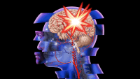
Any patient with a brain injury is at increased risk for cerebral edema. Your ability to recognize cerebral edema early and intervene appropriately can improve patient outcomes. All nurses in medical-surgical and progressive care unit (PCU) settings should be able to recognize signs and symptoms, activate the rapid response team (RRT) as needed, and implement appropriate interventions.
Defining cerebral edema
Cerebral edema is brain swelling following a primary brain injury—an insult to the brain from displacement of its physical structures. Primary brain injury can result from various physiologic or anatomic causes. Because the brain is enclosed within the skull, any changes in pressure or volume can compress brain tissue, leading to damage.
The Monro-Kellie Doctrine is the most common explanation of cerebral volume dynamics involving the brain, cerebrospinal fluid (CSF), and blood within the skull. The doctrine states that to maintain a constant state of volume symmetry, a fixed volume of each of these three components within the skull must be maintained. If the mass or volume of one component increases, one or both of the other components must decrease. For example, when cerebral edema causes increased intracranial pressure (ICP), CSF compensates by moving out of the skull and pooling around the spinal cord. Cerebral edema and increased ICP can lead to permanent brain damage, herniation, or brain death if not controlled in a timely manner.
Classifying cerebral edema
The four major types of cerebral edema are cytotoxic, vasogenic, interstitial, and a combination. (See Comparing cerebral edema types by clicking the PDF icon above.) During the acute injury phase, the specific primary brain injury suggests which type of cerebral edema is most likely to occur, guiding treatment decisions. For example, intracranial hemorrhage can lead to vasogenic edema and the need for osmotic therapy.
Cerebral edema may arise 1 to 18 hours after the initial brain injury and increase steadily until peaking at 48 to 72 hours. Certain variations, such as patient age, brain size, edema location, and comorbidities, can affect symptom type and severity. For example, advanced age correlates directly with brain atrophy and the amount of space available within the skull. Thus, an older adult with cerebral edema may have less severe signs and symptoms because more room is available for the brain to swell. As for edema location, real-estate terms provide a useful analogy: Brain regions above the tentorium are similar to the suburbs, because they have space for growth; regions below the tentorium are urban, where things are compact and there’s little room for growth. These factors can affect the course of treatment.
Patients at risk for developing cerebral edema should be monitored closely in a critical-care setting during the first 48 to 72 hours after the injury and should receive appropriate nursing, medical, and pharmacologic interventions. If a patient is deemed stable and then transferred out of the critical-care setting, med-surg or PCU nurses need to maintain a heightened awareness, be able to identify cerebral edema, and intervene immediately. Conditions that can lead to cerebral edema after the acute 72-hour phase include hydrocephalus and a new brain hemorrhage.
Gauging your patient’s risk
If you’re a med-surg or PCU nurse, you’ll need to assess your patient’s risk for cerebral edema by performing a comprehensive neurologic assessment to establish baseline status and monitor trends. This can help you distinguish preexisting deficits from new ones. You must be able to detect subtle or vague changes in neurologic status to help prevent irreversible damage. A thorough neurologic assessment helps you identify changes quickly and intervene accordingly. Frequently ask yourself, “What’s the worst thing that could happen to my patient today?” This helps you anticipate what your next steps should be in case an emergency arises.
Level of consciousness
The most sensitive and earliest indicator of neurologic deterioration is level of consciousness (LOC). LOC includes arousal and awareness of surroundings and self, which are combined functions of the cerebral hemispheres and brainstem. The slightest LOC change may indicate brain dysfunction. For example, if your patient is alert and oriented during the initial neurologic assessment but later seems lethargic and hard to arouse, you must intervene quickly to decrease the risk of permanent brain damage.
Additional components
Other components of the neurologic assessment include mentation, motor function, sensory function, integrated regulation, and pupillary function.
- Mentation is the ability to focus, comprehend, and remember—functions located mainly in the cerebral cortex. Evaluate the patient’s mentation throughout the head-to-toe assessment by asking questions and asking the patient to follow commands.
- Motor function is regulated by the cerebellar system, voluntary motor system, and lower motor neurons. To assess motor function, determine the patient’s ability to coordinate and execute movements, staying alert for symmetry and strength.
- Sensory function involves the senses, such as touch, sight, and sound, as well as pain. Assessment of the patient’s pain level and location (for instance, headache) can suggest or indicate the location of cerebral edema.
- To assess integrated regulation, check overall function of the nervous system, including vital signs, circulation, elimination, digestion, and emotion. Keep in mind that increased systolic pressure, widened pulse pressure, and slow heart rate are components of Cushing’s triad—a phenomenon that indicates the brain can no longer compensate for cerebral edema and increased ICP.
- Pupillary function changes may indicate neurologic deterioration. Check for pupillary changes that deviate significantly from the patient’s baseline. These may represent a late sign of cerebral edema and increased ICP.
What to do if your patient is at risk
If your assessment determines your patient is at risk for cerebral edema, take appropriate interventions in a timely manner, as described below.
Ensure the ABCs
For a patient with neurologic deterioration, be sure to maintain an adequate airway, breathing, and circulation (ABCs). A patient with a Glasgow Coma Scale score below 8 requires an artificial airway. Maintaining adequate oxygenation and regular breathing patterns optimizes carbon dioxide levels (abnormal levels can compromise cerebral circulation). Closely monitor vital signs (including pulse oximetry), which provide objective data about blood volume circulation. Be aware that vital-sign changes can impair blood circulation and compromise tissue and organ perfusion.
Activate the RRT
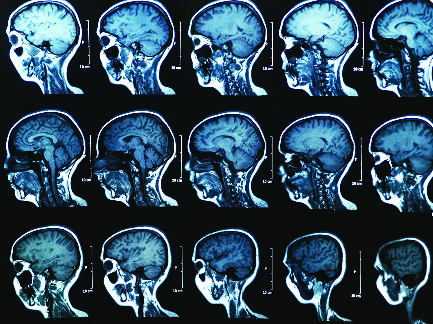
Once you’ve identified neurologic deterioration in your patient, activate the RRT (if your facility has one) and notify the physician right away, to help prevent further deterioration. The RRT provides additional resources to the nurse, patient, and family during a high-acuity situation. If indicated, prepare the patient for diagnostic studies. (See Diagnostic studies for cerebral edema by clicking the PDF icon above.)
Nursing interventions
At the earliest sign of neurologic deterioration or cerebral edema, take the following actions to optimize the patient’s oxygenation, perfusion, and venous drainage and minimize the brain’s metabolic demands.
- Keep the head of the bed elevated 30 or 45 degrees with a neutral head position to promote venous drainage.
- Maintain normal body temperature to minimize metabolic demands and optimize cerebral blood flow. Administer antipyretics, as needed and prescribed.
- Decrease environmental stimuli to minimize the brain’s energy expenditure. Maintain a calm environment with low lighting.
- Monitor fluid and electrolyte levels to help maintain a normal electrolyte balance. Fluid and electrolyte imbalances can worsen cerebral edema.
- Institute seizure precautions in case the patient has a seizure. As prescribed, administer anticonvulsants.
- Institute fall precautions, according to facility policy, to maintain patient safety.
- Provide appropriate pain management to increase patient comfort.
Pharmacologic and surgical interventions
For most causes of cerebral edema, osmotic therapy is the cornerstone of treatment. (See Drugs used to treat cerebral edema by clicking the PDF icon above.) To relieve pressure and prevent further neurologic deterioration, the patient may require surgical intervention, such as a ventriculostomy, craniotomy with debulking, or craniectomy. Indications and the specific procedure are based on the patient’s clinical status.
Left untreated, cerebral edema can lead to permanent and irreversible neurologic damage. Your thorough assessment and timely interventions for patients at risk can help optimize patient outcomes.
Selected references
Hauer EM, Stark D, Staykov D, et al. Early continuous hypertonic saline infusion in patients with severe cerebrovascular disease. Crit Care Med. 2011;39(7):1766-72.
Ho M, Rojas R, Eisenberg RL. Cerebral edema. AJR Am J Roentgenol. 2012;199(3):W258-73.
Inoue K. Caring for the perioperative patient with increased intracranial pressure. AORN J. 2010;91(4):511-8.
Click here for a complete list of references.
Tiffany Niederhauser is a neuroscience clinical nurse specialist at Orlando Regional Medical Center in Orlando, Florida.