The most common sustained cardiac arrhythmia, atrial fibrillation (AF) is reaching epidemic proportions. Not life-threatening in itself, AF is associated with up to twice the mortality risk—even in people without comorbid cardiac conditions.
No matter where you practice, you’re bound to care for patients with AF. More than 2.2 million Americans—and nearly 15% of those older than age 85—experience this arrhythmia. Experts expect the prevalence to increase to 5 million by 2050 as the population ages. This article, which reflects the latest practice guidelines, will enhance your understanding of AF and bring you up to date on current management.
Electrical chaos
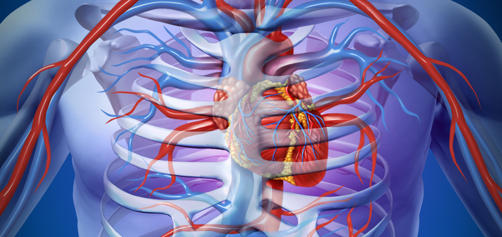
In AF, multiple simultaneous wavelets of electrical activation arise in the atria, causing irregular rapid depolarizations—300 to 600 per minute. This abnormal activity causes the atria to quiver, or fibrillate, and leads to irregular ventricular contractions. The pulse rate becomes irregular, reflecting random conduction of the chaotic electrical impulses. (See Inside the quivering heart in pdf format available by clicking download now.)
Origins of AF
AF can arise as a transient rhythm disturbance in persons with such conditions as thyrotoxicosis or heart failure exacerbation, in those recovering from cardiac or thoracic surgery, and after excessive alcohol intake. Typically, these episodes are paroxysmal (sudden) and may be self-limited. They may even represent a single, isolated incident.
Alternatively, AF may occur as a primary rhythm disturbance in persons without underlying heart disease—a condition termed “lone AF.” In still other cases, AF arises secondary to a cardiac condition or other disease that causes atrial remodeling (dilation and patchy fibrosis within muscle bundles). Such conditions include hypertension, coronary artery disease, valvular disease, pulmonary disease, sleep apnea, and obesity.
Mechanism and triggers
The AF mechanism may be a focal source or it may be more diffuse and involve multiple reentrant circuits. AF triggers include increased sympathetic or parasympathetic tone, premature atrial contractions, bradycardia, and conditions that cause acute atrial stretch (such as pulmonary embolism).
Increased sympathetic tone, as in acute illness, fever, or pneumonia, can trigger AF through excessive atrial depolarization. With increased parasympathetic activity, AF may result from shortening of action potentials, shorter atrial refractory periods, and smaller atrial impulse wavelengths.
How the heart adapts to AF
Within the first 24 hours, the atria adapt to sustained AF through electrical remodeling and by shortening the effective atrial refractory period, which allows for faster rates and more AF.
Nearly two-thirds of AF patients have underlying heart disease that may contribute to structural remodeling—changes in atrial size and structure. Structural remodeling can occur in any setting that promotes atrial stretch, including hypertension, valvular disease, left ventricular hypertrophy, and ventricular dysfunction.
Prolonged rapid rates in AF can lead to significant left atrial enlargement. The larger surface area enables more reentrant circuits to sustain AF, while shortened, heterogeneous atrial refractoriness increases vulnerability to AF. Interstitial fibrosis in muscle bundles may arise in a fibrillating, failing, or aged heart, causing altered cell-to-cell interaction and “zigzag” conduction that perpetuates AF.
Classifying AF
AF is classified as paroxysmal, persistent, or permanent based on its tendency to spontaneously convert to sinus rhythm.
- In paroxysmal AF, episodes come and go, typically last fewer than 24 hours, and convert spontaneously within 7 days.
- In persistent AF, episodes last more than 7 days and require cardioversion with drugs, electrical shock, or both.
- Permanent AF refers to a longstanding episode in which cardioversion fails or no cardioversion effort is made.
Patients may have both parox-ysmal and persistent episodes, but usually their AF is classified by the predominant presentation.
Stroke and other potential sequelae
AF increases the risk of stroke, independent of other risk factors. Blood that pools in the fibrillating atria can lead to clot formation. The clot may break off and end up anywhere in the body; nearly 80% of symptomatic emboli lodge in the brain. In AF patients, the yearly risk of ischemic stroke ranges from 3% to 8%.
Also, many AF patients have concomitant cardiac and other health conditions that raise their overall stroke risk. Adding these risk factors together yields a risk score called CHADS2. (See Scoring your patient’s stroke risk in pdf format available by clicking download now.)
Advancing age influences stroke risk: More than half of AF-associated strokes occur in patients older than age 75. In the Framingham Study, the annual risk of stroke attributable to AF was 23.5% for patients in their 80s, compared to 1.5% for those in their 50s. Patients with AF but no other CHADS2 risk factors (a CHADS2 score of 0) have a fairly low stroke risk.
Tachycardia-induced cardiomyopathy
AF with a rapid ventricular response can lead to left ventricular (LV) enlargement, mitral regurgitation, elevated LV end-diastolic pressure, and a decreased ejection fraction. AF patients with persistently elevated ventricular rates may be unaware of the arrhythmia until they develop heart failure symptoms. They require much the same treatment as in other cases of heart-failure exacerbation—except that controlling the arrhythmia and heart rate may improve or completely reverse the systolic dysfunction.
The rate and duration of tachycardia significantly affect reversibility of tachycardia-induced cardiomyopathy. There’s no particular rate at which such cardiomyopathy occurs; one study suggested it occurs with ventricular rates persistently greater than 130 beats per minute (bpm). Yet persistent rates above 100 bpm also may be sufficient to trigger changes. Therefore, practitioners should suspect tachycardia-induced cardiomyopathy when LV systolic dysfunction occurs in the setting of tachycardia. Early recognition and treatment can help prevent long-term problems.
Assessment and diagnosis
AF doesn’t always cause symptoms. Especially in patients with permanent AF, symptoms typically diminish as palpitations become less noticeable.
But with other AF forms, patients may be extremely symptomatic, complaining of palpitations, shortness of breath, fatigue, dizziness, and even chest pain. These symptoms arise from altered hemodynamic function: With loss of synchronous atrial activity (“atrial kick”), the fibrillating atria no longer contribute to cardiac output, and cardiac output may drop as much as 30%.
With such conditions as diastolic dysfunction, hypertension, restrictive cardiomyopathy, and mitral stenosis, the relative loss in atrial kick is higher and the patient may feel this loss more acutely. Likewise, irregular R-R intervals on an electrocardiogram (ECG) may cause hemodynamic impairment due to variable left ventricular contractility, which contributes to symptoms. Many patients also complain of frequent urination, caused by release of atrial natriuretic peptide (particularly when AF episodes end).
An “irregularly irregular” rhythm
Suspect AF if auscultation reveals an irregular rhythm—but keep in mind that AF is characterized by an irregularly irregular rhythm. This description can help you distinguish AF from sinus rhythm marked by occasional premature contractions (in other words, a rhythm that’s regular for the most part but has irregular moments).
Also remember that patients may have several arrhythmias that can cause an irregular pulse, irregular jugular venous pulsations, or variations in heart-sound intensity. In addition, know that AF may occur alone or in conjunction with atrial flutter or atrial tachycardia; you may have trouble differentiating among these rhythms on auscultation.
ECG confirmation
Although clinical findings may suggest AF, diagnosis requires ECG documentation with at least a single lead recording. In patients with infrequent AF episodes, 24-hour continuous ambulatory monitoring or longer event monitoring (up to 30 days) may be required to document AF.
On the ECG or rhythm strip, AF is diagnosed from characteristic changes in the atrial portion of the electrical complex: Instead of a normal P-QRS combination, P waves are replaced by fibrillatory waves—rapid oscillations of varying amplitudes (usually lower than that of P waves), shape, and periodicity. (See Atrial fibrillation on ECG in pdf format available by clicking download now.) Typically, the QRS complex is normal and narrow, unless the patient has a rate-related bundle-branch block, an accessory pathway, or an underlying distal conduction disease. Sometimes, the ECG shows a characteristic pattern indicating an atrial tachycardia focus firing in short bursts.
A healthy atrioventricular (AV) node can conduct a large number of impulses, so the heart rate may be rapid in AF. But the more often the AV node receives stimulation from the atria, the more slowly it conducts impulses. This decremental property slows conduction enough that the rate doesn’t reflect the 300 to 600 atrial depolarizations taking place each minute.
The heart rate may be near normal in a patient with AV node conduction disease. On the other hand, it may be extremely fast in a patient with an accessory pathway that bypasses the AV node (as in Wolff-Parkinson-White syndrome), because that pathway allows unimpeded conduction.
Diagnostic workup
At the first presentation of AF or in an AF episode with an uncontrolled ventricular response (above 80 bpm), expect the diagnostic workup to include a complete blood count, thyroid panel, hepatic panel, renal function tests, echocardiography, and in many cases a chest X-ray. Test results may show reversible conditions contributing to AF, which can be identified and treated.
Managing atrial fibrillation
The main goals of AF management are to prevent thromboembolic complications and decrease symptoms by controlling the heart rate or restoring normal sinus rhythm.
Preventing thromboembolic complications
The American College of Cardiology/American Heart Association (ACC/AHA) Task Force on Practice Guidelines for AF management (2006) recommends antithrombotic therapy for all patients with AF, except those with lone AF or contraindications for such therapy (such as GI bleeding, previous hemorrhagic stroke, or significant risk of falls).
Warfarin reduces stroke risk by about 70%; aspirin, by about 25%. Drug selection depends on the patient’s CHADS2 score, weighed against the bleeding risk.
In persistent AF, warfarin therapy is recommended starting before sinus rhythm is restored and continuing for at least 4 weeks afterward.
Rate control vs. rhythm control
After addressing anticoagulation, the practitioner determines the best approach for treating AF itself—either controlling heart rate or controlling heart rhythm.
- The goal of rate control is to achieve and maintain a ventricular rate within the normal range.
- The goal of rhythm control is to restore and maintain sinus rhythm.
Selection of approach commonly hinges on the nature of the patient’s AF, expected likelihood that the atrium will sustain a normal rhythm and, perhaps most importantly, the patient’s symptoms.
Rate control. The first step in controlling the heart rate and preventing symptoms from a rapid ventricular response (as well as a possible decrease in heart function) is to administer a beta blocker (such as metoprolol or atenolol), a calcium channel blocker (such as diltiazem or verapamil), digoxin, or a combination of these drugs. In persistent AF, heart rate trends should be assessed through ambulatory monitoring to maintain good rate control (typically 60 to 80 bpm at rest or 90 to 115 bpm with activity).
If adequate rate control proves difficult to achieve, AV node ablation may be done to modify or disconnect electrical conduction between the atria and ventricles and thus prevent rapid ventricular rates.
In some cases, AV node modification slows AF conduction sufficiently. But some patients require complete obliteration of the electrical connection by ablation. In either case, a pacemaker must be inserted to stimulate the heart artificially and initiate a contraction as needed to maintain a healthy heart rate.
Rhythm control. Antiarrhythmic drugs used to be the mainstay of treatment for maintaining sinus rhythm. But the Atrial Fibrillation Follow-up Investigation of Rhythm Management trial showed that for patients with persistent AF, this strategy didn’t reduce deaths more effectively than rate control. However, study subjects eligible for randomization were elderly and had few symptoms, and the antiarrhythmics used had potentially dangerous adverse effects.
In fact, restoring and maintaining sinus rhythm is indicated and attempted in many cases—first presentation of AF, paroxysmal AF, younger patients without structural remodeling, and patients with pronounced AF symptoms. The ACC/AHA Task Force guidelines recommend starting with an appropriate antiarrhythmic, with drug selection based on underlying cardiac disease.
The class IC antiarrhythmics flecainide and propafenone and the class III agents sotalol, dofetilide, and amiodarone are used most frequently, but each has powerful ion-channel-blocking effects and can cause depolarization and repolarization changes. Class IC agents can induce ventricular fibrillation in patients with ischemic damage and shouldn’t be given to those with previous myocardial infarctions or other evidence of coronary artery disease. Dofetilide and sotalol cause QT prolongation and may induce torsades de pointes; their use necessitates hospital admission for loading and 24-hour telemetry monitoring.
If the patient can’t sustain a normal rhythm after trying one or more antiarrhythmics, left-atrial catheter ablation may be done. (See Ending AF with left-atrial catheter ablation in pdf format available by clicking download now.)
Nursing interventions
Provide the following interventions:
- Teach patients with AF about the importance of anticoagulation to reduce stroke risk. Explain that they will be prescribed an anticoagulant drug and must maintain an International Normalized Ratio in the 2.0 to 3.0 range for at least 21 days before conversion to sinus rhythm and for at least 4 weeks afterward. Long-term warfarin therapy is recommended for AF patients with CHADS2 scores of 2 or higher.
- Inform patients they may need to adjust their diet and other medications during warfarin therapy.
- Advise patients that warfarin commonly causes easy bruising and prolonged bleeding, even from minor cuts.
- Provide education about potential adverse drug effects, and stress the importance of getting frequent medical checkups to monitor for such effects.
- Teach patients about possible drug-drug interactions, especially when taking certain antibiotics concomitantly with class III antiarrhythmics. Urge them to tell all prescribers what drugs they’re taking.
- If the patient’s therapeutic regimen changes from medical treatment to catheter ablation, teach the patient and family about this procedure. Inform them that the patient will receive sedation or general anesthesia, and that postprocedure monitoring will include frequent vital sign checks, assessment of groin puncture sites for bleeding or bruising, and telemetry monitoring for arrhythmia.
- After AV node ablation with pacemaker implantation, assess the patient’s incision site and monitor the pocket for hematoma and signs of infection. Also monitor telemetry recordings for potential pacemaker problems.
- Remember that the course of AF therapy may be prolonged, so be sure to provide psychological support. Some patients have symptoms out of proportion to the apparent arrhythmia severity and require frequent reassurance about the nature of the arrhythmia, as well as education about the various treatment approaches available.
Your knowledge of AF and its management can help improve your patients’ quality of life. The more you know about this common arrhythmia, the more prepared you’ll be to provide optimal care, education, and support.
Selected references
Fuster V, Ryden LE, Cannom DS, et al. ACC/AHA/ESC 2006 Guidelines for the Management of Patients with Atrial Fibrillation: a report of the American College of Cardiology/American Heart Association Task Force on Practice Guidelines and the European Society of Cardiology Committee for Practice Guidelines (Writing Committee to Revise the 2001 Guidelines for the Management of Patients With Atrial Fibrillation). Circulation. 2006;114: e257-e354. www.cardiosource.com/guidelines/guidelines/atrial_fib/pdfs/AF_final.pdf. Accessed June 16, 2008.
Go AS, Hylek EM, Phillips KA, et al. Prevalence of diagnosed atrial fibrillation in adults: national implications for rhythm management and stroke prevention: the AnTicoagulation and Risk Factors in Atrial Fibrillation (ATRIA) Study. JAMA. 2001;285:2370-2375. http://jama.ama-assn.org/cgi/content/abstract/285/18/2370. Accessed June 16, 2008.
Hart RG. Atrial fibrillation and stroke prevention. N Engl J Med. 2003;349:1015-1016. http://content.nejm.org/cgi/content/extract/349/11/1015. Accessed June 16, 2008.
Visit www.AmericanNurseToday.com for a complete list of selected references.
Liza A. Prudente is a Nurse Practitioner in the Electrophysiology/Atrial Fibrillation Center at the University of Virginia Health System in Charlottesville. The planners and author of this CNE activity have disclosed no relevant financial relationships with any commercial companies pertaining to this activity.