
Nearly 400,000 cardiac pacemakers and cardioverter-defibrillators are implanted annually in the United States. In some cases, pacemakers are used as a temporary intervention to support a patient through an acute episode. In other situations, as for patients with permanent conditions that require cardiac pacing, pacemakers are implanted surgically.
Whether temporary or permanent, a pacemaker generates an electrical impulse that travels via one or more leadwires, or leads, which in turn stimulate the myocardium to depolarize and initiate a contraction. A pacemaker’s primary function is to keep the ventricles beating at a rate that maintains sufficient blood pressure and perfuses all organs adequately.
Pacemaker components
The main parts of a pacemaker are the pulse generator, which contains the power source (typically a lithium-iodide battery and a small electronic circuit), leads, and electrodes at the terminal ends of the leads. Electrodes may be unipolar or bipolar.
- Bipolar electrodes are located on the same lead. Once the electrode is implanted in the myocardium, the impulse travels to the negative electrode at the tip of the lead, where it stimulates the myocardium. Then the current flows back to the positive electrode to complete the circuit. Compared to unipolar electrodes, bipolar electrodes are less easily affected by outside electrical activity, including skeletal muscle contractions and magnetic fields.
- With a unipolar electrode, a negative electrode is located at the distal tip of the lead and the positive electrode is located at the pulse generator. Unipolar electrodes are more sensitive to myocardial electrical activity.
Single-chamber vs. dual-chamber pacemakers
Single-chamber pacemakers are used primarily to pace the ventricle when the patient’s underlying rhythm is atrial fibrillation or another atrial arrhythmia. Temporary single-chamber pacemakers most commonly are used in emergencies when temporary pacing is required. If the patient has an atrial arrhythmia, the lead for a single-chamber pacemaker is placed in the right ventricle. If the patient has intact atrioventricular (AV) conduction with a slow heart rate, the lead is placed in the right atrium.
Dual-chamber pacemakers—the most commonly implanted pacemaker type—have two leads. The pulse generator is implanted in the chest wall; one lead is implanted in the right atrium and the other in the right ventricle. The pacemaker then maintains synchrony between the atria and ventricles. Dual-chamber pacemakers are used to treat AV node dysfunction, acquired AV blocks, and advanced second- and third-degree heart blocks in adults, as well as chronic bifascicular blocks.
Pacemaker codes
Many clinicians use the North American Society for Pacing and Electrophysiology/British Pacing and Electrophysiology Group (NASPE/BPEG) generic code for Antibradycardia, Adaptive-Rate Pacing, and Multisite Pacing. This system describes pacemaker and automatic implantable cardioverter-defibrillator settings. (See NASPE/BPEG generic code for pacemakers by clicking the PDF icon above.)
Temporary pacemakers
Temporary pacing is indicated when the heart rate must be increased urgently or as a preventive measure—for instance, in postoperative cardiac surgery patients. Right coronary artery myocardial infarction (MI) commonly causes bradycardic arrhythmias due to lack of blood flow to the sinoatrial node. In a large percentage of individuals, the right coronary artery also perfuses the AV node. Many drugs, including diltiazem, digitalis, and beta blockers, slow the heart rate and in toxic doses may cause symptomatic bradycardia. These conditions may call for temporary pacing.
Normally, all types of temporary pacing are demand pacing, in which the pacemaker delivers electrical current only when the heart’s intrinsic rate falls below the preset rate. The three types of temporary pacing are transcutaneous, transvenous, and epicardial.
Transcutaneous pacing
Transcutaneous pacing is used in the most urgent situations. The American Heart Association’s Advanced Cardiac Life Support Guidelines recommend noninvasive transcutaneous pacing (NTP) for hemodynamically unstable bradycardia. When used for symptomatic bradycardia, NTP is a Class I recommendation—always acceptable, unquestionably safe, and definitely useful. NTP also is recommended to ready a patient for pacing if needed. For example, if a patient has the potential to develop a heart block (as after an MI), transcutaneous pacing pads are placed on the patient so pacing can begin if symptomatic bradycardia develops.
Today, most defibrillators have a temporary pacing function. With NTP, one large pacing pad is placed on the patient’s chest wall and another on the back; both are attached to the defibrillator set in the pacing mode. (See Pad placement for temporary transcutaneous pacing by clicking the PDF icon above.) When the patient’s heart rate falls below the programmed rate, the NTP fires or paces the myocardium through the chest wall. The amount of milliamperes (mA) required to stimulate the myocardium through the chest wall is higher than when an electrode is placed directly on the myocardium; for NTP, up to 200 mA may be used. Because NTP may be uncomfortable for patients who are awake, sedation and analgesia should be used if possible.
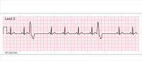
With NTP, electrocardiograph (ECG) leads are attached to the patient to sense a QRS complex, which represents ventricular depolarization. Like other ventricular demand pacemakers (which may be temporary or permanent), an NTP fires only when the intrinsic heart rate falls below a set rate. When it senses a QRS complex, it inhibits firing. (See Evaluating pacemaker rhythm strips by clicking the PDF icon above.)
Transvenous pacing
Transvenous pacing is used after the recommended 12 hours of continuous or 24 hours of intermittent transcutaneous pacing have elapsed. It’s also used as a bridge to permanent pacemaker implantation. A leadwire is guided through the vascular system to the myocardium and attached to a cable that connects to the pulse generator.
Transvenous pacing is relatively comfortable for the patient. The pacemaker rate can be set at a low of 40 to 60 paced beats; the pacemaker fires when the rate drops below the set rate. In some models, the rate can be set for up to 110 beats/minute. Transvenous pacemakers have built-in sensing to identify ventricular depolarization.
A temporary transvenous pacemaker may be set between 0 and 20 mA; the average setting is 10 mA. The pulse generator’s sensitivity settings
allow the generator to “see” intrinsic myocardial activity more clearly. Sensitivity should be set so the pulse generator can “see” intrinsic P and QRS waves, which appear on the ECG and are measured in millivolts (mV). An initial ventricular sensitivity setting of
2 to 5 mV is the recommended starting point for ventricular pacing. If sensitivity is set too high, electrical activity won’t be “seen” and the pacemaker will fire—a phenomenon called undersensing or failure to sense. If sensitivity is set too low, the pacemaker may recognize electrical artifacts, such as muscle movements, and will fail to fire; this is known as oversensing.
In some patients, two pacing leads may be placed—one in the atrium and the other in the ventricle. When programmed for AV sequential pacing, the pacemaker is used to increase cardiac output by maintaining the heart’s natural cycle (atrial contraction followed by ventricular contraction). Additional settings for AV sequential pacing include sensitivity for atrial pacing and the AV interval (the interval between atrial pacing and ventricular pacing). For atrial pacing, the average setting is 0.5 mV (which is lower than ventricular sensitivity because the P wave is smaller than the QRS complex). In a normal heart, the AV interval is about 200 milliseconds. Pacemaker AV intervals can be set to mimic the patient’s normal PR interval.
Epicardial pacing
Epicardial pacing is used only with cardiac surgery patients. Common postoperative complications of cardiac valve surgery and coronary artery bypass surgery include episodic sinus bradycardia, second- and third-degree heart block, and asystole. During the intraoperative period, epicardial pacing wires are attached to the epicardium, passed through the chest wall, and attached to a cable connected to the pacemaker’s pulse generator. Epicardial pacemaker settings are similar to transvenous pacemaker settings.
An endothelial sheath may form around the tips of the pacing wires, affecting capture and sensing. When this happens, the mA setting may have to be raised to achieve capture and the sensitivity setting may have to be lowered to achieve sensing. The patient’s fluid and electrolyte status also influences the ability to pace and sense properly. Pacing of both the atria and ventricles (dual-chamber pacing) maintains cardiac synchrony and optimizes preload and cardiac output.
Permanent pacemakers
Permanent pacemakers are implanted during a short surgical procedure. The pulse generator is placed in a subcutaneous pocket created in the chest wall—usually on the upper left part of the chest below the clavicle. Then the pulse generator is attached to leads, which are threaded through the vascular system to the heart and implanted into the myocardial wall. The atrial electrode is implanted in the right atrium near the coronary
sinus; the ventricular electrode, near the right ventricular apex. Currently, most implanted pacemakers are dual-chamber pacemakers; about one-third have cardioversion-defibrillation settings. Implanted cardioverter-defibrillator (ICD) use is approved as a first-line treatment for patients at risk for ventricular tachycardia or ventricular fibrillation.
Permanent pacemakers are used to treat various bradycardic arrhythmias. (See Permanent pacemaker indications by clicking the PDF icon above.) Usually, patients are evaluated for bradycardia based on ECG records from 24-hour or Holter monitoring.
Biventricular pacing
Also called cardiac resynchronization therapy, biventricular pacing is indicated for patients with heart failure, dilated cardiomyopathy, prolonged ventricular conduction, or a reduced ejection fraction. Asynchronous activation of the heart chambers (indicated by a QRS duration greater than 0.13 seconds) is linked to worsening heart failure and increased morbidity and mortality. A biventricular pacemaker has three leads—one for the atrium and one for each ventricle. Biventricular pacing causes synchronous contraction of both ventricles and improves cardiac output.
End-of-life concerns
In terminally ill patients and those who’ve opted to forgo further resuscitative efforts, pacemakers with ICD functionality may interfere with the natural process of dying by continuing to function and delivering shocks. As the end of the patient’s life approaches, the patient and family should discuss with healthcare providers whether the ICD function should be discontinued.
If you’re caring for a monitored hospital patient with a pacemaker, remember that the ECG will continue to show pacing spikes and possible electrical activity without a pulse. This could cause confusion in family members at the bedside, so the healthcare team should consider monitoring the patient
remotely.
What the future may hold
Experts predict that permanent pacemakers of the future will be much smaller—about the size of a large grain of rice—and will be implanted intravascularly, not surgically. What’s more, the pulse generator will be seated directly into the myocardium without leads. For children, this means fewer surgeries to replace leads in a growing body. For adults, benefits include the possibility of fewer lead fractures and less lead displacement.
Each year, the number of patients who receive permanent pacemakers grows. Although pacemaker technology is changing constantly, the goal of therapy remains the same—to sustain the heart rate and improve AV synchrony, thus optimizing cardiac output and improving the patient’s quality of life.
Selected references
Bernstein AD, Daubert JC, Fletcher RD, et al. The Revised NASPE/BPEG generic code for antibradycardia, adaptive-rate, and multisite pacing. North American Society of Pacing and Electrophysiology/British Pacing and Electrophysiology Group. Pacing Clin Electrophysiol. 2002 (Feb);25(2):260-4.
Chow WC, Buxton AE. Implantable Cardiac Pacemakers and Defibrillators: All You Wanted to Know. Malden, MA: Blackwell Publishing, Inc; 2006.
ECG Interpretation: An Incredibly Visual! Pocket Guide. Philadelphia, PA: Lippincott Williams & Wilkins; 2009.
Epstein AE, DiMarco JP, Ellenbogen KA, et al. ACC/AHA/HRS 2008 Guidelines for Device-Based Therapy of Cardiac Rhythm Abnormalities: a report of the American College of Cardiology/American Heart Association Task Force on Practice Guidelines (Writing Committee to Revise the ACC/AHA/NASPE 2002 Guidelines Update for Implantation of Cardiac Pacemakers and Antiarrhythmic Devices) developed in collaboration with the American Association of Thoracic Surgery and Society of Thoracic Surgeons. J Am Coll Cardiol. 2008 May 27;51(21);e1-62. Erratum in: J Am Coll Cardiol. 2009 Apr 21;53(16):1473. J Am Coll Cardiol. 2009 Jan 6;53(1):147.
Field JM, Hazinski MF, Sayre MR, et al. 2010 American Heart Association Guidelines for Cardiopulmonary Resuscitation and Emergency Cardiovascular Care Science. Circulation. 2010;122(suppl 3):S640-656. http://circ.ahajournals.org/content/122/18_suppl_3.toc. Accessed January 30, 2012.
Indications for use of non-invasive transcutaneous pacing. Resuscitation Central. http://www.resuscitationcentral.com/pacing/indications. Accessed January 30, 2012.
Jackson A. An overview of permanent cardiac pacing. Nurs Stand. 2010 Nov 24-30; 25(12):47-57.
Runge MS, Stouffer GA, Patterson C. Netter’s Cardiology (Netter Clinical Science). 2nd ed. Philadelphia, PA: Saunders; 2010.
Russo JE. Original research: deactivation of ICDs at the end of life: a systemic review of clinical practices and provider and patient attitudes. Am J Nurs. 2011 Oct:111(10):26-35.
Scheibly K. Systematic assessment of basic pacemaker function. AACN Adv Crit Care. 2010 Jul-Sep:21(3);322-8.
The eleven most implanted medical devices in America. 24/7 Wall St. Wire. July 18, 2011. http://247wallst.com/2011/07/18/the-eleven-most-implanted-medical-devices-in-america/2/. Accessed January 30, 2012.
Visit www.AmericanNurseToday.com for a complete list of references.
Nicolette C. Mininni is an advanced practice nurse in critical care at UPMC Shadyside in Pittsburgh, Pennsylvania.


4 Comments.
My comment is the same as the others above. This site is really a poor example of user friendliness. All attempts have been useless. I have been a member of ANA since 1969 and find this lack of consideration to members grounds for discontinuing membership and PAC contributions.
completed a test. Could not enter my ANA number anywhere to get discount and was then unable to print certificate of completion. Would not use this program again if this difficut.
I have the same problem. Logging in is technically much too difficult. I get blank screens right and left. Nothing should not be this hard.
I have had difficulty acquiring CEUs from your website for the past three years. As a result, I always have to send the info in by mail. Is there someone who can help me. I have three e-mail addresses. I have tried all of them as well as all of the passwords that I have used. Yesterday and today I have tried changing my passord numerous times and am unable to do so. I have tried re-registering and have been told that I already exist in your files – whichch I already knew. What recourse?