Enteral feedings deliver nourishment through a tube directly into the GI tract. They’re ordered for patients with a functioning GI tract who can’t ingest enough nutrition orally to meet their needs. The feeding tube may stay in place as briefly as a few days or permanently, until the patient’s death. (See Indications for enteral feeding.)
This article discusses types of enteral feeding tubes, methods, and formulas. It also reviews enteral feeding complications and describes related nursing care.
Defining malnutrition
People experiencing the physiologic stress of illness may have increased metabolic demands with reduced capacity to take in nutrition. Prolonged calorie restriction can lead to malnutrition. According to the Academy of Nutrition and Dietetics and the American Society for Parenteral and Enteral Nutrition (A.S.P.E.N.), patients with at least two of the following criteria are malnourished:
Enteral feeding: Indications, complications, and nursing care
Top 10 care essentials for ventilator patients
• insufficient energy intake
• weight loss
• muscle mass loss
• subcutaneous fat loss
• localized or generalized fluid accumulation that may mask weight loss
• diminished functional status as measured by handgrip strength.
Malnourished patients with inadequate caloric and protein intake may suffer emaciation, poor healing, and pressure injuries. In severe cases, they may develop osteopenia, osteomalacia, osteoporosis, muscle weakness, increased fracture risk, polyneuropathy, paresthesias, confusion, dementia, and pancytopenia. Some also may have low albumin and prealbumin levels, which can cause fluid to pool in a localized or generalized distribution. But before blaming malnutrition for abnormal albumin or prealbumin levels, clinicians must consider such factors as persistent inflammation and hepatic or renal impairment.
Types of enteral feeding tubes
The practitioner selects the type of feeding tube based on the specific enteral formula the patient requires and the anticipated duration of enteral feeding. The two main types of feeding tubes are prepyloric and postpyloric.
• Prepyloric tubes end in the stomach above the pyloric sphincter. They’re preferred for intermittent feeding and to allow gastric absorption.
• Postpyloric tubes end beyond the pyloric sphincter in the jejunum. They’re indicated for patients with gastroparesis, acute pancreatitis, gastric outlet stenosis, hyper emesis (including gravida), recurrent aspiration, tracheoesophageal fistula, and stenosis with gastroenterostomy. Postpyloric feedings must be administered on a continuous basis. (See Comparing enteral feeding tubes.)
Enteral feeding formulas
Entering feeding formulas fall into several general categories, such as polymeric formulas, feeding modules, elemental, and specialized or disease-specific formulas. Practitioners choose the formula that bests meet the patient’s individual needs. Nutritional demands vary with age, weight, height, current nutritional status, laboratory values, and activity level. Also, enteral feeding requirements may vary even within similar groups of patients, such as those with renal dysfunction or liver failure.
To calculate the correct volume to deliver, practitioners consider the total protein, fat, and carbohydrates needed to restore the patient’s health. Some dietitians start with a basic formula of 25- to 35-cal/kg and adjust it to the patient’s condition. Certain medical conditions create a higher metabolic demand, necessitating increased feeding volume.
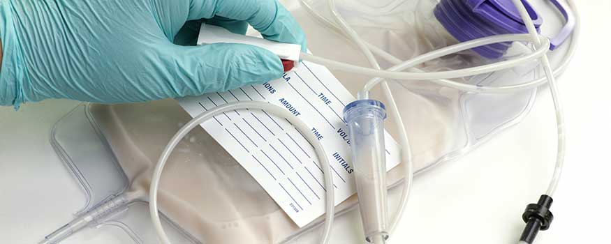
Enteral formulas can be administered using either an open or a closed (ready-to-use) system and can be delivered through several methods. (See Understanding enteral feeding systems and methods.)
Complications of enteral feeding
Patients with feeding tubes are at risk for such complications as aspiration, tube malpositioning or dislodgment, refeeding syndrome, medication-related complications, fluid imbalance, insertion-site infection, and agitation. To identify these problems, thoroughly assess the patient before tube feeding begins and monitor closely during feedings. (For information on insertion-site infection and agitation, see Other enteral-feeding complications.)
Aspiration
Gastrostomy (G) tube feedings can cause pulmonary aspiration. Multiple factors contribute to aspiration, including recent hemorrhagic stroke, high gastric residual volume (GRV), high bolus feeding volumes, supine positioning, and conditions that affect the esophageal sphincters (such as an indwelling endotracheal or tracheostomy tube with dysfunction of the upper esophageal sphincter and a nasogastric or an enteral tube traversing both esophageal sphincters).
Studies show that patients who received tube feedings of 500 to 1,500 mL/day didn’t have a higher aspiration risk than those fed lower daily volumes; even some who received low volumes aspirated. However, relatively fast feeding rates with volumes exceeding 1,500 mL/day did place patients at higher risk for aspiration.
To help reduce risk, monitor GRV every 4 hours (or according to protocol) in patients receiving continuous tube feedings. A.S.P.E.N. and the Society of Critical Care Medicine guidelines for critically ill patients advise against halting tube feedings for GRVs below 500 mL unless the patient has other signs and symptoms of intolerance. Sometimes, healthcare providers order withholding of tube feedings at lower GRVs because of specific risk factors.
If you find tube feeding contents in the patient’s mouth during oral care, assume the presence of reflux, which increases aspiration risk. To help prevent this problem, keep the head of the bed elevated 30 degrees or higher when possible.
During patient transport or when placing the head of the bed flat for patient repositioning, turn the tube feeding off, especially if the patient has a high aspiration risk. However, be aware that no conclusive evidence shows that pausing tube feeding during repositioning reduces aspiration risk for patients with high GRVs.
Tube malpositioning or dislodgment
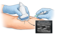
During initial placement, the feeding tube may be positioned improperly. To prevent this problem, the tube should be placed by experienced personnel and its position confirmed radiographically. After initial placement, the tube may become fully or partially dislodged, causing such problems as bleeding, tracheal or parenchymal perforation, and GI tract perforation.
To help prevent malpositioning and dislodgment, verify feeding tube integrity at the beginning of each shift. Be aware that verbal patients with dislodged tubes may complain of new-onset pain at or near the insertion site of a percutaneous endoscopic gastrostomy (PEG) tube, G tube, gastric-jejunal (GJ) tube, or J tube. Nonverbal patients may respond with vital-sign changes (such as increased blood pressure or heart rate), increased agitation, and restlessness.
Refeeding syndrome
Patients with sustained malnutrition are at risk for refeeding syndrome—the body’s reaction to digestion after depleted electrolytes shift from the serum to the intracellular space. This syndrome may trigger life-threatening arrhythmias and multisystemic dysfunction. It occurs when a depleted metabolic system with little to no mineral reserve (for instance, from vitamin B deficiency) becomes exhausted by the body’s increased demands to process proteins and produce glycogen. An insulin response to reintroducing nutrition causes an anaerobic state, as the body can’t meet the demand for oxygen and other resources needed to metabolize nutrients. Serum electrolytes then move into the intracellular space to help satisfy the higher demands, resulting in acute electrolyte abnormalities.
In patients with long-term malnutrition, monitor for intolerance at the onset of enteral feedings by checking heart rate and rhythm and electrolyte levels. Although refeeding syndrome incidence is low, failure to recognize the sudden drop in potassium and magnesium levels can have catastrophic consequences.
To reduce the risk of refeeding syndrome in patients with vitamin and mineral deficiencies, supplements may be ordered for parenteral administration before enteral feedings begin. Refer to specific guidelines based on total energy needs and specific micronutrient deficiencies; thiamine and other B-vitamin deficiencies are the most pressing ones to address before initiating enteral feeding. As the tube-feeding goal rate is achieved, taper micronutrient supplement dosages as indicated.
Fluid imbalance
Most patients need supplemental free-water flushes to maintain adequate hydration; on average, they need 30 mL/kg of water per day, given either as free-water flushes or I.V. hydration.
If a free-water flush is ordered, calculate its volume by subtracting the volume of water in the feeding formula from the patient’s total daily requirement; then divide the remaining volume over a regular routine of tube flushing. Before and after medication administration, flush the tube with about 30 mL of fluid or more, depending on drug characteristics. Note: Be aware that some patients are at high risk for fluid overload and depend on a concentrated feeding formula to meet dietary needs.
Medication-related complications
Until recently, clinicians assumed diarrhea in patients receiving enteral feedings stemmed from malabsorption and feeding intolerance. But more recent research points to medications, especially those high in sorbitol, as the main culprit. So be sure to rule out medications as the cause of diarrhea before looking for other causes, including malabsorption and rapid delivery rates.
The sorbitol content of certain premade liquid drugs (such as potassium chloride, acetaminophen, and theophylline) can cause a rapid fluid shift into the intestines, leading to hyperosmolarity and diarrhea. This effect increases when sorbitolbased liquid medications are given through a J tube. (Gastric acid in the stomach acts as a buffer to medications and reduces osmolarity of fluid entering the small intestine.) Consider a pharmacy consult for patient who experience diarrhea while receiving multiple sorbitol-based drugs. Changing the administration time as appropriate or switching to a non-sorbitol-based alternative may relieve diarrhea without necessitating feeding-rate adjustment.
Medications administered through a feeding tube also may cause clogging, especially if they’re crushed. Don’t give medications that must be crushed through a J tube, because the clogging risk is greater than with a G tube. Take additional precautions with medications linked to a higher clogging risk, including psyllium, ciprofloxacin suspension, sevelamer, and potassium chloride tablets that can be dissolved in water.
Know that tube replacement due to clogging is costly and subjects the patient to anesthesia. To help prevent clogging, maintain proper tube maintenance and flushing. For instance, massage potential clots in the tube, irrigate with warm water, administer alkalinized enzymes as ordered, and use a manual declogger (such as the Bionix DeClogger® or CorPak Clog Zapper®) if needed.
Be aware that some medications must be given on an empty stomach to ensure effective absorption, including phenytoin, carbama zepine, alendronate, carbidopa levodopa, and levothyroxine. You may need to withhold tube feedings for 1 to 2 hours before and after administering these medications. For a patient with a GJ tube, as long as medications are given through the gastric port, you needn’t withhold feedings from the jejunal port; follow pharmacy guidelines. Keep in mind that patients receiving multiple drugs may have absorption problems due to extended withholding of feedings, causing dehydration and malnutrition.
Nursing care
When beginning enteral feedings, monitor the patient for feeding tolerance. Assess the abdomen by auscultating for bowel sounds and palpating for rigidity, distention, and tenderness. Know that patients who complain of fullness or nausea after a feeding starts may have higher a GRV.
On an ongoing basis, monitor patients for gastric distention, nausea, bloating, and vomiting. Stop the infusion and notify the provider if the patient experiences acute abdominal pain, abdominal rigidity, or vomiting.
Multidisciplinary approach to patient care
Successful management of the patient’s nutritional needs requires a team approach. Each discipline is responsible for managing and monitoring the patient’s physiologic and psychological needs. Caloric requirements calculated by a dietitian must be ordered by a healthcare provider and delivered and monitored by a nurse. (However, some states permit dietitians to initiate nutritional interventions.) Nursing assistants can help with patient positioning and comfort care as well as behavioral monitoring. Consult additional specialists, such as a wound ostomy nurse, about the risk of pressure injuries compounded by malnutrition or dehydration.
Keep the goal of care in mind. For terminally ill patients, palliative care specialists can help evaluate the benefits and risks of continuing enteral feeding and help clinicians navigate ethical issues, such as whether to continue enteral feedings and other life-prolonging measures. They can also help manage symptoms and make suggestions based on the patient’s or family’s goal of care.
Future of enteral feedings
Enteral feedings have the potential to advance patient care. For example, enteral formulas specific to the patient’s condition and fluid requirements are now available. Also, trials currently are underway in critical care units to study the use of feeding tubes with magnetic components at the end, which could allow confirmation of correct tube placement with a magnet instead of radiography. As technology progresses, enteral feeding efficiency will progress as well. For the best outcomes, healthcare providers must work as a team to treat the patient holistically.
The authors work at Halifax Health in Daytona Beach, Florida. Amanda Houston is a professional development education specialist at Halifax Health in Daytona Beach, Florida. Paul Fuldauer is a clinical nutritional coordinator.
Selected references
Agre P, Brown P, Stone K. Tube feeding troubleshooting guide. The Oley Foundation. March 2016.
American Geriatrics Society Ethics Committee and Clinical Practice and Models of Care Committee. American Geriatrics Society feeding tubes in advanced dementia position statement. J Am Geriatr Soc. 2014;62(8):1590-3.
Arabi YM, Aldawood AS, Haddad SH, et al; PermiT Trial Group. Permissive underfeeding or standard enteral feeding in critically ill adults. N Engl J Med. 2015;372(25):2398-408.
Bankhead R, Boullata J, Brantley S, et al; A.S.P.E.N. Board of Directors. Enteral nutrition practice recommendations. JPEN J Parenter Enteral Nutr. 2009;33(2):122-67.
Camilleri M, Parkman HP, Shafi MA, et al. Clinical guideline: management of gastroparesis. Am J Gastroenterol. 2013;108(1):18-37.
Chen S, Xian W, Cheng S, et al. Risk of regurgitation and aspiration in patients infused with different volumes of enteral nutrition. Asia Pac J Clin Nutr. 2015;24(2):212-8.
Dolman RC, Conradie C, Lombard MJ, Nienaber A, Wicks M. Case study: Nutritional management of a patient at high risk of developing refeeding syndrome. S Afr J Clin Nutr. 2015;28(3):140-5.
Jensen GL, Compher C, Sullivan DH, Mullin GE. Recognizing malnutrition in adults: definitions and characteristics, screening, assessment, and team approach. JPEN J Parenter Enteral Nutr. 2013;37(6):802-7.
John BK, Bullock M, Brenner L, McGaw C, Scolapio JS. Nutrition in the elderly. Frequently asked questions. Am J Gastroenterol. 2013;108(8):1252-66.
Jongbloed F, Ijzermans JN, de Bruin RW. Permissive underfeeding or standard enteral feeding in critical illness. N Engl J Med. 2015;373(12):1175.
Martino R, Martin RE, Black S. Dysphagia after stroke and its management. CMAJ. 2012;184(10):1127-8.
McClave SP, Taylor BE, Martindale RG, et al; Society of Critical Care Medicine, American Society for Parenteral and Enteral Nutrition. Guidelines for the provision and assessment of nutrition support therapy in the adult critically ill patient: Society of Critical Care Medicine (SCCM) and American Society for Parenteral and Enteral Nutrition (A.S.P.E.N.). JPEN J Parenter Enteral Nutr. 2016;40(2):159-211.
Ojo O. Enteral feeding tubes: not perfect but necessary. Br J Nurs. 2015;24(18):910.
Powers J, Fischer MH, Ziemba-Davis M, Brown J, Phillips DM. Elimination of radiographic confirmation for small-bowel feeding tubes in critical care. Am J Crit Care. 2013;22(6):521-7.
Romano MM. Medicines and tube feeding formula. 2014 Oley Conference; Tube Feeding Workshop Presentation. June 24, 2014.
Seminerio J, O’Keefe SJ. Jejunal feeding in patients with pancreatitis. Nutr Clin Pract. 2014;29(3):283-6.
Schwartz L. Choosing the right tube for you. LifelineLetter. The Oley Foundation. January/February 2014.
Walmsley RS. Refeeding syndrome: screening, incidence, and treatment during parenteral nutrition. J Gastroenterol Hepatol. 2013;28 Suppl 4:113-7.
White JV, Guenter P, Jensen G, Malone A, Schofield M; Academy of Nutrition and Dietetics Malnutrition Work Group; A.S.P.E.N. Malnutrition Task Force; A.S.P.E.N. Board of Directors. Consensus statement of the Academy of Nutrition and Dietetics/American Society for Parenteral and Enteral Nutrition: characteristics recommended for the identification and documentation of adult malnutrition (undernutrition). J Acad Nutr Diet. 2012;112(5):730-8.


7 Comments.
Very informative
Thank you for your comment!
Question: If one uses a 1000cc feeding bag, is it ok to add more formula to the bag if it wasn’t all used during the previous feeding? May I please be added to your mailing list?
It’s good to learn here. Please tabulate nursing actions and rationale in administering tube feeding.
Thanks
I would love to be added to your mailing list.
I work in acute care in Sanford, Florida. HCA facility.
Thank you!
Belen
I cannot find the cne’s I bought this morning
Very interesting article.