Family medical history reveals risk for sudden cardiac death.
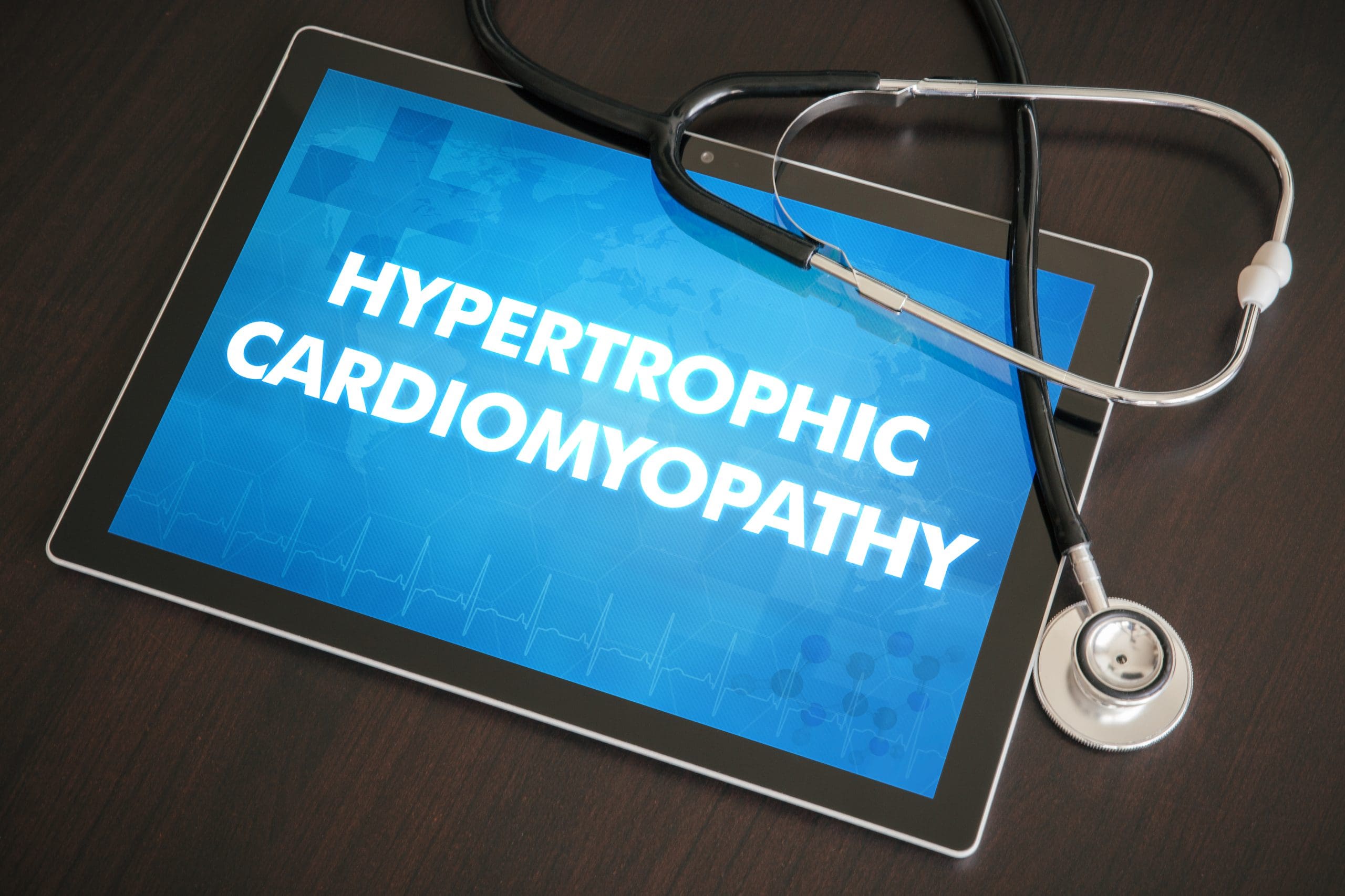
- Hypertrophic cardiomyopathy (HCM) is the number one cause of sudden cardiac death in young people.
- HCM usually results from an autosomal dominant genetic mutation.
- An extensive multigenerational family history and review of systems can help reduce the risk of sudden cardiac death and lead to prompt diagnosis and treatment.
John Benson*, a 45-year-old man, arrives in the ED with chest discomfort, shortness of breath, and palpitations that have worsened. Andre, the ED nurse, notes Mr. Benson’s pallor and Mr. Benson explains he was working outdoors and felt faint.
History and assessment
Mr. Benson’s father died suddenly of a heart issue at the age of 38. He notes that he’s previously been told that he has a heart murmur.
Pulmonary hypertension: Consider the “zebra”
An overview of arrhythmogenic cardiomyopathy
During the physical examination, Andre notes that the patient’s resting HR is 110 bpm, RR 24 breaths per minute, BP 90/40 mmHg, and O2 saturation 97. Cardiovascular examination reveals a harsh systolic ejection murmur at the left sternal border and a weak second heart sound. The patient has dry mouth and poor skin turgor.
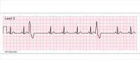
Andre recognizes these signs and symptoms as possible warning signs for myocardial infarction, or other serious heart condition, and dehydration. He follows the organization’s cardiac protocol and obtains an ECG, which shows sinus tachycardia with possible left atrial enlargement and left ventricular hypertrophy as evidenced by ST segment depression and T wave inversion in the left-sided leads. Andre starts a 20-gauge I.V. and draws blood for a CBC, CMP, and troponin. He also initiates lactated Ringer’s at 50 mL/hr and oxygen at 2L via nasal canula. The first troponin in the series is negative.
The ED provider orders a stat echocardiogram, which shows asymmetric septal hypertrophy with a maximum thickness of 21 mm (normal is 8 mm to 14 mm), and systolic anterior motion of the mitral valve, resulting in significant obstruction of the left ventricular outflow track (gradient of 90 mmHg at rest) and moderate mitral regurgitation. The left atrium is dilated (43 mm) with normal LV systolic function (ejection fraction 68%). Cardiac MRI confirms the ultrasound findings and reveals minor myocardial fibrosis. The ED provider diagnoses Mr. Benson with hypertrophic obstructive cardiomyopathy, the most common type of hypertrophic cardiomyopathy. After consultation with a cardiologist, the provider orders metoprolol at 50 mg once daily for 14 days, increasing the dose every two weeks by 50 mg. to achieve a 200 mg. daily dose goal. Mr. Benson is then discharged home.
Outcome
Mr. Benson’s heart doesn’t respond to medical therapy and he requires invasive treatment. A septal myectomy to remove part of the hypertrophied septal wall, relieves the obstruction. Mr. Benson has an uneventful recovery and is discharged home on beta-blockers. Genetic testing reveals a common hypertrophic cardiomyopathy gene variant myosin-binding protein C.
The cardiologist advises Mr. Benson to avoid strenuous physical activity and to take nebivolol 5 mg once a day in the morning, follow-up visits and echocardiograhy. Genetic counseling and cardiac testing are recommended for his children and blood relatives.
Education and follow-up
Hypertrophic cardiomyopathy comprises two subtypes: obstructive and nonobstructive. The condition’s clinical presentation ranges from asymptomatic to severe heart failure. It’s the most common cause of sudden cardiac death in young people and most commonly is diagnosed in the second or third decade of life.
Mr. Benson’s case highlights the importance of completing a multigenerational family history. This history and a physical assessment can reduce the risk of sudden cardiac death.
*Names are fictitious.
Erik P. Southard is a professor and interim associate director of academics–nursing at Indiana State University School of Nursing in Terre Haute.
American Nurse Journal. 2024; 19(3). Doi: 10.51256/ANJ032430
References
Affronti A, Pruna-Guillen R, Sandoval E, Pereda D, Alcocer J, Castellà M, Quintana E. Surgery for hypertrophic obstructive cardiomyopathy: Comprehensive LVOT management beyond septal myectomy. J Clin Med. 2021;10(19):4397. doi:10.3390/jcm10194397
Hodges K, Rivas CG, Aguilera J, et al. Surgical management of left ventricular outflow tract obstruction in a specialized hypertrophic obstructive cardiomyopathy center. J Thorac Cardiovasc Surg. 2019;157(6):2289-99. doi:10.1016/j.jtcvs.2018.11.148
Hoss S, Habib M, Silver J, et al. Genetic testing for diagnosis of hypertrophic cardiomyopathy mimics: Yield and clinical significance. Circ Genom Precis Med. 2020;13(2):e002748. doi:10.1161/CIRCGEN.119.002748
Mandes L, Rosca M, Ciupercă D, Popescu BA. The role of echocardiography for diagnosis and prognostic stratification in hypertrophic cardiomyopathy. J Echocardiogr. 2020;18(3):137-48. doi:10.1007/s12574-020-00467-9
Maron BJ, Desai MY, Nishimura RA, et al. Management of hypertrophic cardiomyopathy: JACC state-of-the-art review. J Am Coll Cardiol. 2022;79(4):390-414. doi:10.1016/j.jacc.2021.11.021
Key words: cardiomyopathy, hypertrophic cardiomyopathy, sudden cardiac death, chest pain


1 Comment. Leave new
Become an operating room nurse you love the job